Calcium »
PDB 3ecj-3eqd »
3ela »
Calcium in PDB 3ela: Crystal Structure of Active Site Inhibited Coagulation Factor Viia Mutant in Complex with Soluble Tissue Factor
Enzymatic activity of Crystal Structure of Active Site Inhibited Coagulation Factor Viia Mutant in Complex with Soluble Tissue Factor
All present enzymatic activity of Crystal Structure of Active Site Inhibited Coagulation Factor Viia Mutant in Complex with Soluble Tissue Factor:
3.4.21.21;
3.4.21.21;
Protein crystallography data
The structure of Crystal Structure of Active Site Inhibited Coagulation Factor Viia Mutant in Complex with Soluble Tissue Factor, PDB code: 3ela
was solved by
J.R.Bjelke,
M.Fodje,
L.A.Svensson,
with X-Ray Crystallography technique. A brief refinement statistics is given in the table below:
| Resolution Low / High (Å) | 29.19 / 2.20 |
| Space group | P 1 21 1 |
| Cell size a, b, c (Å), α, β, γ (°) | 78.361, 68.558, 78.817, 90.00, 90.22, 90.00 |
| R / Rfree (%) | 23.3 / 29.4 |
Calcium Binding Sites:
The binding sites of Calcium atom in the Crystal Structure of Active Site Inhibited Coagulation Factor Viia Mutant in Complex with Soluble Tissue Factor
(pdb code 3ela). This binding sites where shown within
5.0 Angstroms radius around Calcium atom.
In total only one binding site of Calcium was determined in the Crystal Structure of Active Site Inhibited Coagulation Factor Viia Mutant in Complex with Soluble Tissue Factor, PDB code: 3ela:
In total only one binding site of Calcium was determined in the Crystal Structure of Active Site Inhibited Coagulation Factor Viia Mutant in Complex with Soluble Tissue Factor, PDB code: 3ela:
Calcium binding site 1 out of 1 in 3ela
Go back to
Calcium binding site 1 out
of 1 in the Crystal Structure of Active Site Inhibited Coagulation Factor Viia Mutant in Complex with Soluble Tissue Factor

Mono view
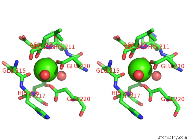
Stereo pair view
Mono view
Stereo pair view
A full contact list of Calcium with other atoms in the Ca binding
site number 1 of Crystal Structure of Active Site Inhibited Coagulation Factor Viia Mutant in Complex with Soluble Tissue Factor within 5.0Å range:
|
Reference:
J.R.Bjelke,
O.H.Olsen,
M.Fodje,
L.A.Svensson,
S.Bang,
G.Bolt,
B.B.Kragelund,
E.Persson.
Mechanism of the CA2+-Induced Enhancement of the Intrinsic Factor Viia Activity J.Biol.Chem. V. 283 25863 2008.
ISSN: ISSN 0021-9258
PubMed: 18640965
DOI: 10.1074/JBC.M800841200
Page generated: Sat Jul 13 09:30:39 2024
ISSN: ISSN 0021-9258
PubMed: 18640965
DOI: 10.1074/JBC.M800841200
Last articles
Zn in 9J0NZn in 9J0O
Zn in 9J0P
Zn in 9FJX
Zn in 9EKB
Zn in 9C0F
Zn in 9CAH
Zn in 9CH0
Zn in 9CH3
Zn in 9CH1