Calcium »
PDB 3eqf-3f45 »
3erc »
Calcium in PDB 3erc: Crystal Structure of the Heterodimeric Vaccinia Virus Mrna Polyadenylate Polymerase with Three Fragments of Rna and 3'-Deoxy Atp
Enzymatic activity of Crystal Structure of the Heterodimeric Vaccinia Virus Mrna Polyadenylate Polymerase with Three Fragments of Rna and 3'-Deoxy Atp
All present enzymatic activity of Crystal Structure of the Heterodimeric Vaccinia Virus Mrna Polyadenylate Polymerase with Three Fragments of Rna and 3'-Deoxy Atp:
2.1.1.57; 2.7.7.19;
2.1.1.57; 2.7.7.19;
Protein crystallography data
The structure of Crystal Structure of the Heterodimeric Vaccinia Virus Mrna Polyadenylate Polymerase with Three Fragments of Rna and 3'-Deoxy Atp, PDB code: 3erc
was solved by
C.Li,
H.Li,
S.Zhou,
T.L.Poulos,
P.D.Gershon,
with X-Ray Crystallography technique. A brief refinement statistics is given in the table below:
| Resolution Low / High (Å) | 38.55 / 3.21 |
| Space group | P 1 |
| Cell size a, b, c (Å), α, β, γ (°) | 69.925, 77.153, 108.030, 89.52, 73.45, 63.76 |
| R / Rfree (%) | 24.5 / 30.9 |
Calcium Binding Sites:
The binding sites of Calcium atom in the Crystal Structure of the Heterodimeric Vaccinia Virus Mrna Polyadenylate Polymerase with Three Fragments of Rna and 3'-Deoxy Atp
(pdb code 3erc). This binding sites where shown within
5.0 Angstroms radius around Calcium atom.
In total 4 binding sites of Calcium where determined in the Crystal Structure of the Heterodimeric Vaccinia Virus Mrna Polyadenylate Polymerase with Three Fragments of Rna and 3'-Deoxy Atp, PDB code: 3erc:
Jump to Calcium binding site number: 1; 2; 3; 4;
In total 4 binding sites of Calcium where determined in the Crystal Structure of the Heterodimeric Vaccinia Virus Mrna Polyadenylate Polymerase with Three Fragments of Rna and 3'-Deoxy Atp, PDB code: 3erc:
Jump to Calcium binding site number: 1; 2; 3; 4;
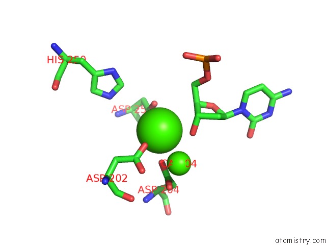
Calcium binding site 1 out of 4 in 3erc
Go back to
Calcium binding site 1 out
of 4 in the Crystal Structure of the Heterodimeric Vaccinia Virus Mrna Polyadenylate Polymerase with Three Fragments of Rna and 3'-Deoxy Atp
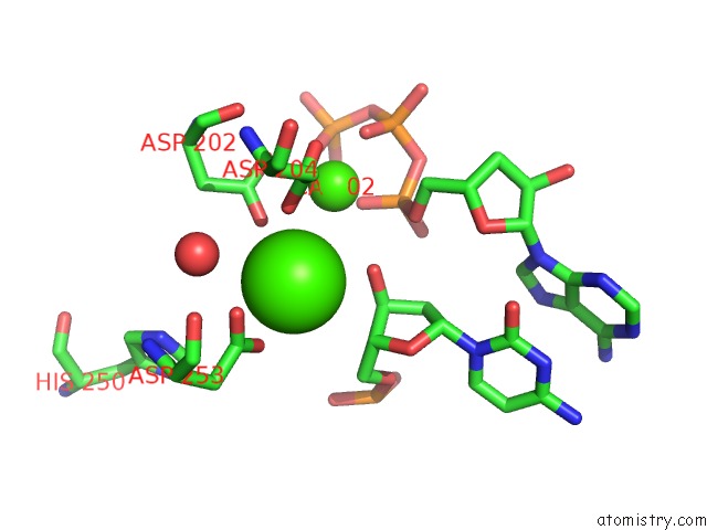
Mono view
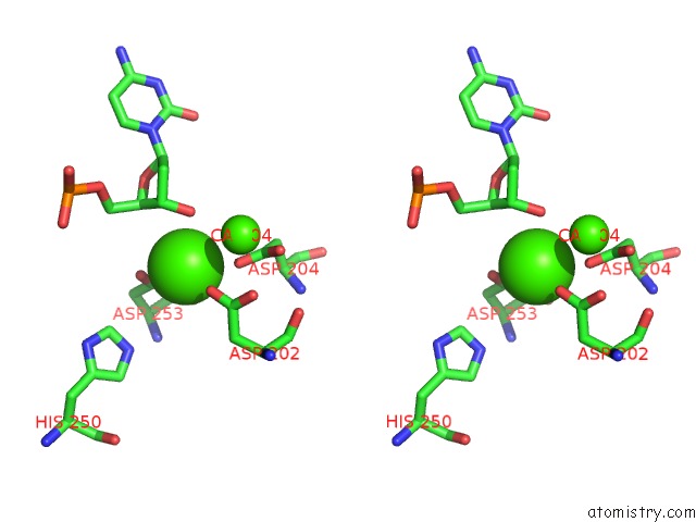
Stereo pair view
Mono view
Stereo pair view
A full contact list of Calcium with other atoms in the Ca binding
site number 1 of Crystal Structure of the Heterodimeric Vaccinia Virus Mrna Polyadenylate Polymerase with Three Fragments of Rna and 3'-Deoxy Atp within 5.0Å range:
|
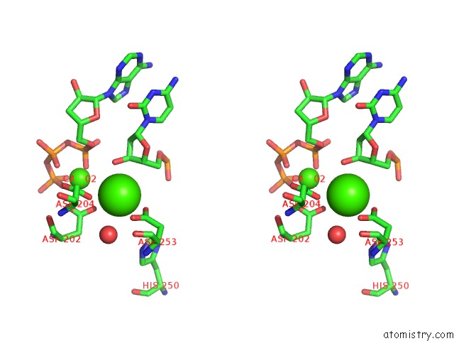
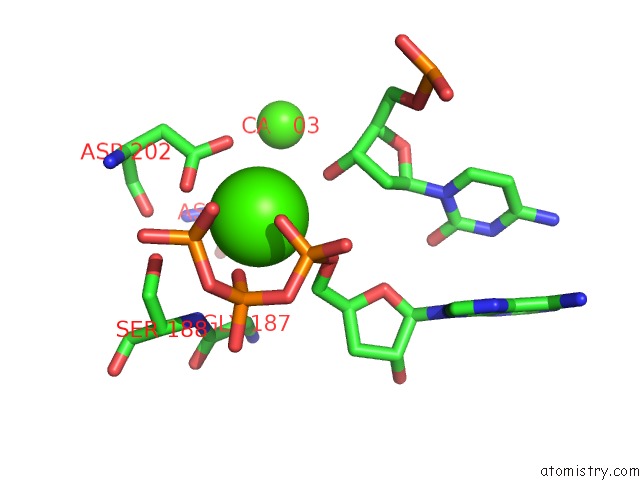
Calcium binding site 2 out of 4 in 3erc
Go back to
Calcium binding site 2 out
of 4 in the Crystal Structure of the Heterodimeric Vaccinia Virus Mrna Polyadenylate Polymerase with Three Fragments of Rna and 3'-Deoxy Atp
Mono view
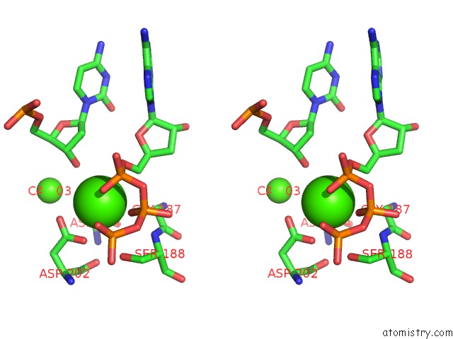
Stereo pair view
Mono view
Stereo pair view
A full contact list of Calcium with other atoms in the Ca binding
site number 2 of Crystal Structure of the Heterodimeric Vaccinia Virus Mrna Polyadenylate Polymerase with Three Fragments of Rna and 3'-Deoxy Atp within 5.0Å range:
|
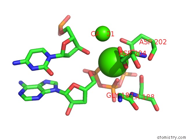
Calcium binding site 3 out of 4 in 3erc
Go back to
Calcium binding site 3 out
of 4 in the Crystal Structure of the Heterodimeric Vaccinia Virus Mrna Polyadenylate Polymerase with Three Fragments of Rna and 3'-Deoxy Atp
Mono view
Stereo pair view
Mono view
Stereo pair view
A full contact list of Calcium with other atoms in the Ca binding
site number 3 of Crystal Structure of the Heterodimeric Vaccinia Virus Mrna Polyadenylate Polymerase with Three Fragments of Rna and 3'-Deoxy Atp within 5.0Å range:
|
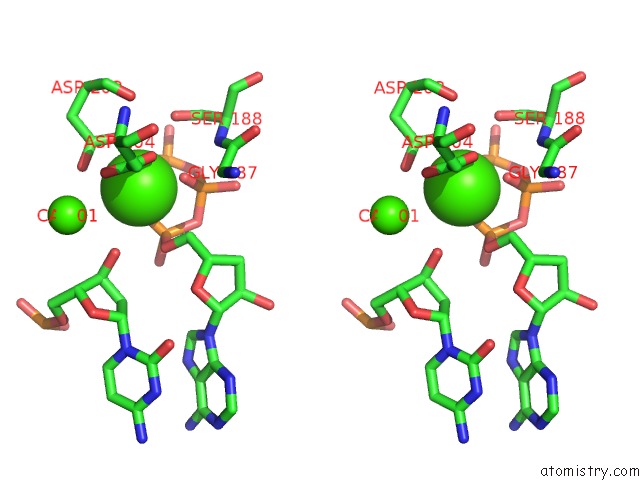
Calcium binding site 4 out of 4 in 3erc
Go back to
Calcium binding site 4 out
of 4 in the Crystal Structure of the Heterodimeric Vaccinia Virus Mrna Polyadenylate Polymerase with Three Fragments of Rna and 3'-Deoxy Atp
Mono view
Stereo pair view
Mono view
Stereo pair view
A full contact list of Calcium with other atoms in the Ca binding
site number 4 of Crystal Structure of the Heterodimeric Vaccinia Virus Mrna Polyadenylate Polymerase with Three Fragments of Rna and 3'-Deoxy Atp within 5.0Å range:
|
Reference:
C.Li,
H.Li,
S.Zhou,
E.Sun,
J.Yoshizawa,
T.L.Poulos,
P.D.Gershon.
Polymerase Translocation with Respect to Single-Stranded Nucleic Acid: Looping or Wrapping of Primer Around A Poly(A) Polymerase Structure V. 17 680 2009.
ISSN: ISSN 0969-2126
PubMed: 19446524
DOI: 10.1016/J.STR.2009.03.012
Page generated: Tue Jul 8 11:59:24 2025
ISSN: ISSN 0969-2126
PubMed: 19446524
DOI: 10.1016/J.STR.2009.03.012
Last articles
Ca in 7G6ICa in 7G6H
Ca in 7G6F
Ca in 7G6G
Ca in 7G6E
Ca in 7G6D
Ca in 7G6B
Ca in 7G6C
Ca in 7G6A
Ca in 7G69