Calcium »
PDB 3eqf-3f45 »
3etr »
Calcium in PDB 3etr: Crystal Structure of Xanthine Oxidase in Complex with Lumazine
Protein crystallography data
The structure of Crystal Structure of Xanthine Oxidase in Complex with Lumazine, PDB code: 3etr
was solved by
J.M.Pauff,
H.Cao,
R.Hille,
with X-Ray Crystallography technique. A brief refinement statistics is given in the table below:
| Resolution Low / High (Å) | 50.00 / 2.20 |
| Space group | P 1 21 1 |
| Cell size a, b, c (Å), α, β, γ (°) | 133.190, 73.491, 146.505, 90.00, 98.68, 90.00 |
| R / Rfree (%) | 19.7 / 26.7 |
Other elements in 3etr:
The structure of Crystal Structure of Xanthine Oxidase in Complex with Lumazine also contains other interesting chemical elements:
| Molybdenum | (Mo) | 2 atoms |
| Iron | (Fe) | 8 atoms |
Calcium Binding Sites:
The binding sites of Calcium atom in the Crystal Structure of Xanthine Oxidase in Complex with Lumazine
(pdb code 3etr). This binding sites where shown within
5.0 Angstroms radius around Calcium atom.
In total only one binding site of Calcium was determined in the Crystal Structure of Xanthine Oxidase in Complex with Lumazine, PDB code: 3etr:
In total only one binding site of Calcium was determined in the Crystal Structure of Xanthine Oxidase in Complex with Lumazine, PDB code: 3etr:
Calcium binding site 1 out of 1 in 3etr
Go back to
Calcium binding site 1 out
of 1 in the Crystal Structure of Xanthine Oxidase in Complex with Lumazine
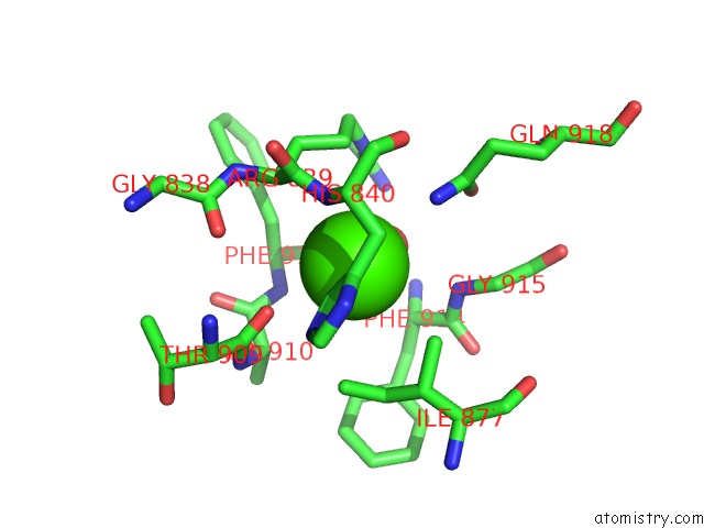
Mono view
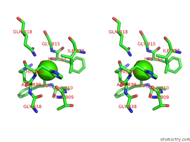
Stereo pair view
Mono view
Stereo pair view
A full contact list of Calcium with other atoms in the Ca binding
site number 1 of Crystal Structure of Xanthine Oxidase in Complex with Lumazine within 5.0Å range:
|
Reference:
J.M.Pauff,
H.Cao,
R.Hille.
Substrate Orientation and Catalysis at the Molybdenum Site in Xanthine Oxidase: Crystal Structures in Complex with Xanthine and Lumazine. J.Biol.Chem. V. 284 8760 2009.
ISSN: ISSN 0021-9258
PubMed: 19109252
DOI: 10.1074/JBC.M804517200
Page generated: Sat Jul 13 09:35:38 2024
ISSN: ISSN 0021-9258
PubMed: 19109252
DOI: 10.1074/JBC.M804517200
Last articles
Zn in 9J0NZn in 9J0O
Zn in 9J0P
Zn in 9FJX
Zn in 9EKB
Zn in 9C0F
Zn in 9CAH
Zn in 9CH0
Zn in 9CH3
Zn in 9CH1