Calcium »
PDB 3f5v-3fib »
3f9p »
Calcium in PDB 3f9p: Crystal Structure of Myeloperoxidase From Human Leukocytes
Enzymatic activity of Crystal Structure of Myeloperoxidase From Human Leukocytes
All present enzymatic activity of Crystal Structure of Myeloperoxidase From Human Leukocytes:
1.11.1.7;
1.11.1.7;
Protein crystallography data
The structure of Crystal Structure of Myeloperoxidase From Human Leukocytes, PDB code: 3f9p
was solved by
X.Carpena,
I.Fita,
C.Obinger,
with X-Ray Crystallography technique. A brief refinement statistics is given in the table below:
| Resolution Low / High (Å) | 20.00 / 2.93 |
| Space group | P 43 21 2 |
| Cell size a, b, c (Å), α, β, γ (°) | 110.740, 110.740, 255.333, 90.00, 90.00, 90.00 |
| R / Rfree (%) | 23.6 / 25.7 |
Other elements in 3f9p:
The structure of Crystal Structure of Myeloperoxidase From Human Leukocytes also contains other interesting chemical elements:
| Iron | (Fe) | 2 atoms |
| Chlorine | (Cl) | 2 atoms |
Calcium Binding Sites:
The binding sites of Calcium atom in the Crystal Structure of Myeloperoxidase From Human Leukocytes
(pdb code 3f9p). This binding sites where shown within
5.0 Angstroms radius around Calcium atom.
In total 2 binding sites of Calcium where determined in the Crystal Structure of Myeloperoxidase From Human Leukocytes, PDB code: 3f9p:
Jump to Calcium binding site number: 1; 2;
In total 2 binding sites of Calcium where determined in the Crystal Structure of Myeloperoxidase From Human Leukocytes, PDB code: 3f9p:
Jump to Calcium binding site number: 1; 2;
Calcium binding site 1 out of 2 in 3f9p
Go back to
Calcium binding site 1 out
of 2 in the Crystal Structure of Myeloperoxidase From Human Leukocytes

Mono view
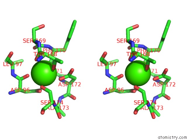
Stereo pair view
Mono view
Stereo pair view
A full contact list of Calcium with other atoms in the Ca binding
site number 1 of Crystal Structure of Myeloperoxidase From Human Leukocytes within 5.0Å range:
|
Calcium binding site 2 out of 2 in 3f9p
Go back to
Calcium binding site 2 out
of 2 in the Crystal Structure of Myeloperoxidase From Human Leukocytes
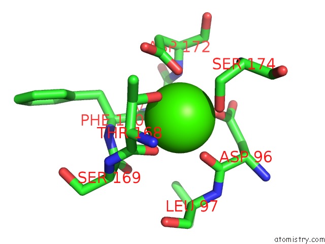
Mono view

Stereo pair view
Mono view
Stereo pair view
A full contact list of Calcium with other atoms in the Ca binding
site number 2 of Crystal Structure of Myeloperoxidase From Human Leukocytes within 5.0Å range:
|
Reference:
X.Carpena,
P.Vidossich,
K.Schroettner,
B.M.Calisto,
S.Banerjee,
J.Stampler,
M.Soudi,
P.G.Furtmuller,
C.Rovira,
I.Fita,
C.Obinger.
Essential Role of Proximal Histidine-Asparagine Interaction in Mammalian Peroxidases. J.Biol.Chem. V. 284 25929 2009.
ISSN: ISSN 0021-9258
PubMed: 19608745
DOI: 10.1074/JBC.M109.002154
Page generated: Sat Jul 13 09:43:30 2024
ISSN: ISSN 0021-9258
PubMed: 19608745
DOI: 10.1074/JBC.M109.002154
Last articles
Zn in 9J0NZn in 9J0O
Zn in 9J0P
Zn in 9FJX
Zn in 9EKB
Zn in 9C0F
Zn in 9CAH
Zn in 9CH0
Zn in 9CH3
Zn in 9CH1