Calcium »
PDB 3f5v-3fib »
3fax »
Calcium in PDB 3fax: The Crystal Structure of Gbs Pullulanase Sap in Complex with Maltotetraose
Protein crystallography data
The structure of The Crystal Structure of Gbs Pullulanase Sap in Complex with Maltotetraose, PDB code: 3fax
was solved by
L.J.Gourlay,
with X-Ray Crystallography technique. A brief refinement statistics is given in the table below:
| Resolution Low / High (Å) | 40.00 / 2.40 |
| Space group | P 21 21 21 |
| Cell size a, b, c (Å), α, β, γ (°) | 48.214, 102.862, 171.690, 90.00, 90.00, 90.00 |
| R / Rfree (%) | 22.1 / 28.3 |
Other elements in 3fax:
The structure of The Crystal Structure of Gbs Pullulanase Sap in Complex with Maltotetraose also contains other interesting chemical elements:
| Chlorine | (Cl) | 1 atom |
Calcium Binding Sites:
The binding sites of Calcium atom in the The Crystal Structure of Gbs Pullulanase Sap in Complex with Maltotetraose
(pdb code 3fax). This binding sites where shown within
5.0 Angstroms radius around Calcium atom.
In total 4 binding sites of Calcium where determined in the The Crystal Structure of Gbs Pullulanase Sap in Complex with Maltotetraose, PDB code: 3fax:
Jump to Calcium binding site number: 1; 2; 3; 4;
In total 4 binding sites of Calcium where determined in the The Crystal Structure of Gbs Pullulanase Sap in Complex with Maltotetraose, PDB code: 3fax:
Jump to Calcium binding site number: 1; 2; 3; 4;
Calcium binding site 1 out of 4 in 3fax
Go back to
Calcium binding site 1 out
of 4 in the The Crystal Structure of Gbs Pullulanase Sap in Complex with Maltotetraose

Mono view
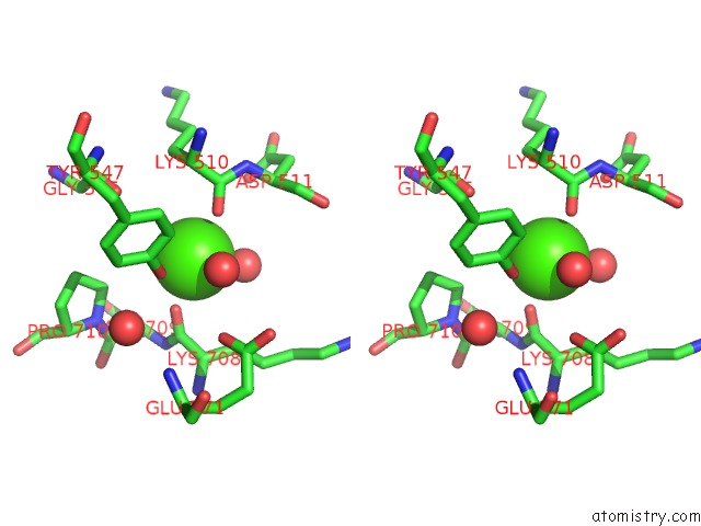
Stereo pair view
Mono view
Stereo pair view
A full contact list of Calcium with other atoms in the Ca binding
site number 1 of The Crystal Structure of Gbs Pullulanase Sap in Complex with Maltotetraose within 5.0Å range:
|
Calcium binding site 2 out of 4 in 3fax
Go back to
Calcium binding site 2 out
of 4 in the The Crystal Structure of Gbs Pullulanase Sap in Complex with Maltotetraose
Mono view

Stereo pair view
Mono view
Stereo pair view
A full contact list of Calcium with other atoms in the Ca binding
site number 2 of The Crystal Structure of Gbs Pullulanase Sap in Complex with Maltotetraose within 5.0Å range:
|
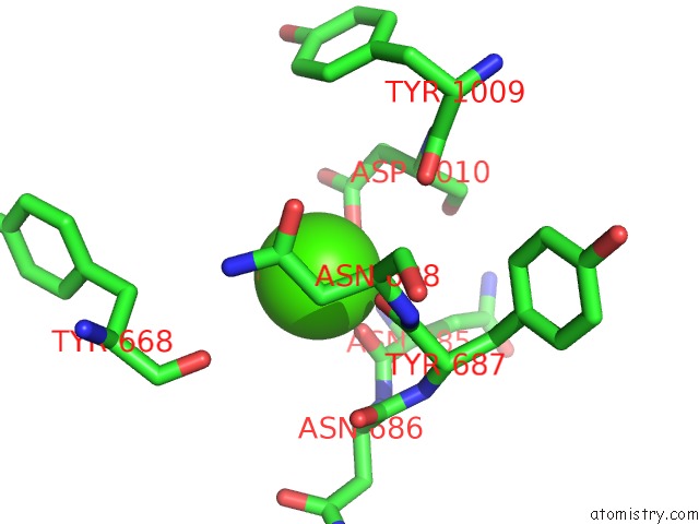
Calcium binding site 3 out of 4 in 3fax
Go back to
Calcium binding site 3 out
of 4 in the The Crystal Structure of Gbs Pullulanase Sap in Complex with Maltotetraose

Mono view

Stereo pair view
Mono view
Stereo pair view
A full contact list of Calcium with other atoms in the Ca binding
site number 3 of The Crystal Structure of Gbs Pullulanase Sap in Complex with Maltotetraose within 5.0Å range:
|
Calcium binding site 4 out of 4 in 3fax
Go back to
Calcium binding site 4 out
of 4 in the The Crystal Structure of Gbs Pullulanase Sap in Complex with Maltotetraose

Mono view

Stereo pair view
Mono view
Stereo pair view
A full contact list of Calcium with other atoms in the Ca binding
site number 4 of The Crystal Structure of Gbs Pullulanase Sap in Complex with Maltotetraose within 5.0Å range:
|
Reference:
L.J.Gourlay,
I.Santi,
A.Pezzicoli,
G.Grandi,
M.Soriani,
M.Bolognesi.
Group B Streptococcus Pullulanase Crystal Structures in the Context of A Novel Strategy For Vaccine Development J.Bacteriol. V. 191 3544 2009.
ISSN: ISSN 0021-9193
PubMed: 19329633
DOI: 10.1128/JB.01755-08
Page generated: Sat Jul 13 09:44:06 2024
ISSN: ISSN 0021-9193
PubMed: 19329633
DOI: 10.1128/JB.01755-08
Last articles
Zn in 9J0NZn in 9J0O
Zn in 9J0P
Zn in 9FJX
Zn in 9EKB
Zn in 9C0F
Zn in 9CAH
Zn in 9CH0
Zn in 9CH3
Zn in 9CH1