Calcium »
PDB 3fil-3fye »
3fl8 »
Calcium in PDB 3fl8: Crystal Structure of B. Anthracis Dihydrofolate Reductase (Dhfr) with RAB1, A Tmp-Dihydrophthalazine Derivative
Enzymatic activity of Crystal Structure of B. Anthracis Dihydrofolate Reductase (Dhfr) with RAB1, A Tmp-Dihydrophthalazine Derivative
All present enzymatic activity of Crystal Structure of B. Anthracis Dihydrofolate Reductase (Dhfr) with RAB1, A Tmp-Dihydrophthalazine Derivative:
1.5.1.3;
1.5.1.3;
Protein crystallography data
The structure of Crystal Structure of B. Anthracis Dihydrofolate Reductase (Dhfr) with RAB1, A Tmp-Dihydrophthalazine Derivative, PDB code: 3fl8
was solved by
C.R.Bourne,
W.W.Barrow,
with X-Ray Crystallography technique. A brief refinement statistics is given in the table below:
| Resolution Low / High (Å) | 24.97 / 2.29 |
| Space group | P 21 21 21 |
| Cell size a, b, c (Å), α, β, γ (°) | 68.178, 135.920, 168.652, 90.00, 90.00, 90.00 |
| R / Rfree (%) | 20.6 / 25.4 |
Calcium Binding Sites:
The binding sites of Calcium atom in the Crystal Structure of B. Anthracis Dihydrofolate Reductase (Dhfr) with RAB1, A Tmp-Dihydrophthalazine Derivative
(pdb code 3fl8). This binding sites where shown within
5.0 Angstroms radius around Calcium atom.
In total 8 binding sites of Calcium where determined in the Crystal Structure of B. Anthracis Dihydrofolate Reductase (Dhfr) with RAB1, A Tmp-Dihydrophthalazine Derivative, PDB code: 3fl8:
Jump to Calcium binding site number: 1; 2; 3; 4; 5; 6; 7; 8;
In total 8 binding sites of Calcium where determined in the Crystal Structure of B. Anthracis Dihydrofolate Reductase (Dhfr) with RAB1, A Tmp-Dihydrophthalazine Derivative, PDB code: 3fl8:
Jump to Calcium binding site number: 1; 2; 3; 4; 5; 6; 7; 8;
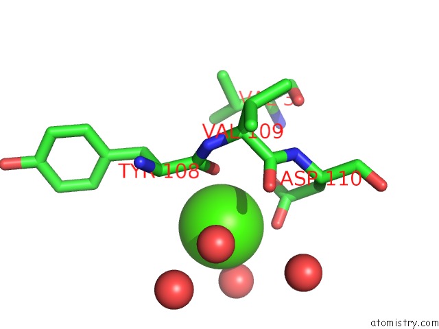
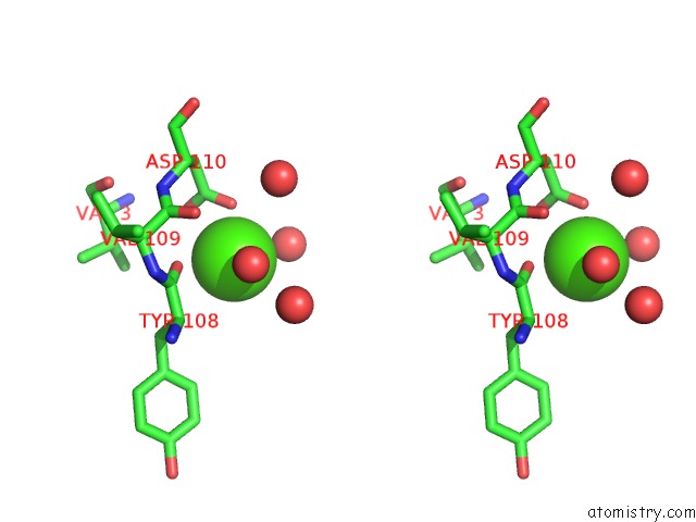
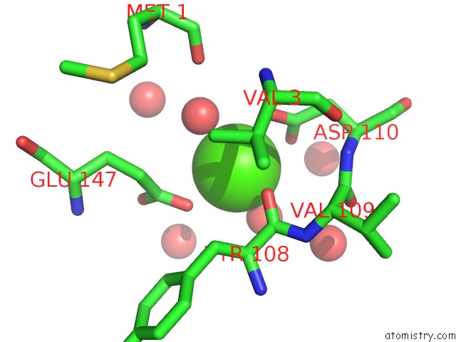
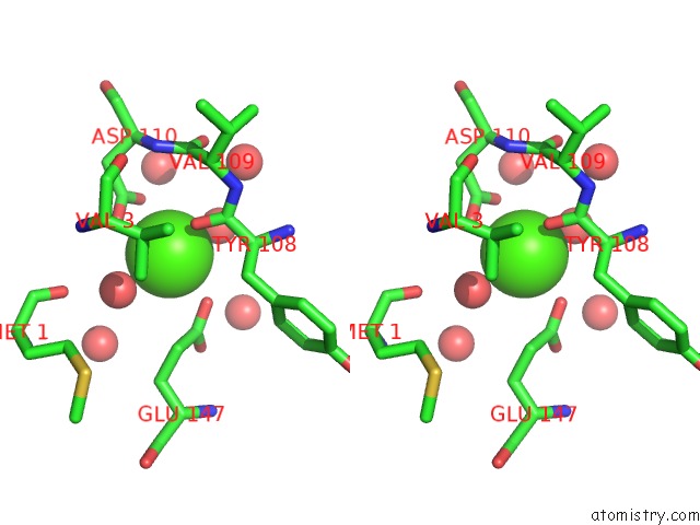
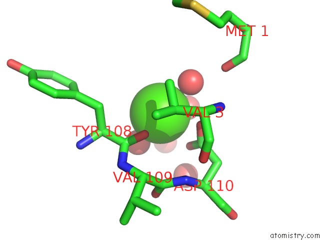
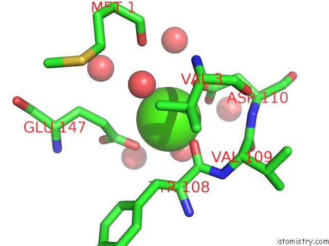
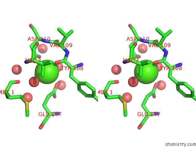
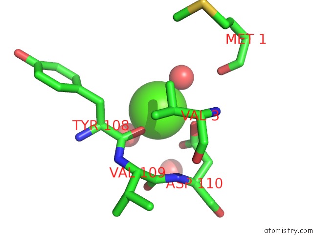
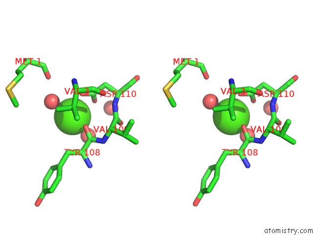
Calcium binding site 1 out of 8 in 3fl8
Go back to
Calcium binding site 1 out
of 8 in the Crystal Structure of B. Anthracis Dihydrofolate Reductase (Dhfr) with RAB1, A Tmp-Dihydrophthalazine Derivative
Mono view
Stereo pair view
Mono view
Stereo pair view
A full contact list of Calcium with other atoms in the Ca binding
site number 1 of Crystal Structure of B. Anthracis Dihydrofolate Reductase (Dhfr) with RAB1, A Tmp-Dihydrophthalazine Derivative within 5.0Å range:
|
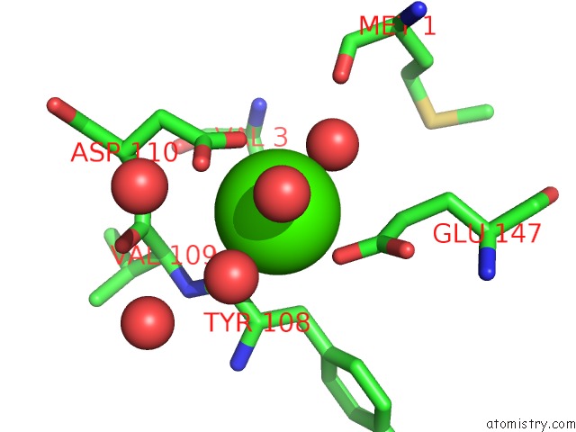
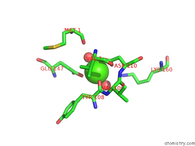
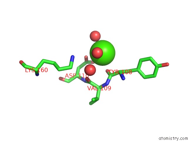
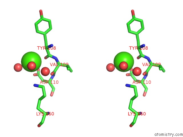
Calcium binding site 2 out of 8 in 3fl8
Go back to
Calcium binding site 2 out
of 8 in the Crystal Structure of B. Anthracis Dihydrofolate Reductase (Dhfr) with RAB1, A Tmp-Dihydrophthalazine Derivative
Mono view
Stereo pair view
Mono view
Stereo pair view
A full contact list of Calcium with other atoms in the Ca binding
site number 2 of Crystal Structure of B. Anthracis Dihydrofolate Reductase (Dhfr) with RAB1, A Tmp-Dihydrophthalazine Derivative within 5.0Å range:
|
Calcium binding site 3 out of 8 in 3fl8
Go back to
Calcium binding site 3 out
of 8 in the Crystal Structure of B. Anthracis Dihydrofolate Reductase (Dhfr) with RAB1, A Tmp-Dihydrophthalazine Derivative
Mono view
Stereo pair view
Mono view
Stereo pair view
A full contact list of Calcium with other atoms in the Ca binding
site number 3 of Crystal Structure of B. Anthracis Dihydrofolate Reductase (Dhfr) with RAB1, A Tmp-Dihydrophthalazine Derivative within 5.0Å range:
|
Calcium binding site 4 out of 8 in 3fl8
Go back to
Calcium binding site 4 out
of 8 in the Crystal Structure of B. Anthracis Dihydrofolate Reductase (Dhfr) with RAB1, A Tmp-Dihydrophthalazine Derivative
Mono view
Stereo pair view
Mono view
Stereo pair view
A full contact list of Calcium with other atoms in the Ca binding
site number 4 of Crystal Structure of B. Anthracis Dihydrofolate Reductase (Dhfr) with RAB1, A Tmp-Dihydrophthalazine Derivative within 5.0Å range:
|
Calcium binding site 5 out of 8 in 3fl8
Go back to
Calcium binding site 5 out
of 8 in the Crystal Structure of B. Anthracis Dihydrofolate Reductase (Dhfr) with RAB1, A Tmp-Dihydrophthalazine Derivative
Mono view
Stereo pair view
Mono view
Stereo pair view
A full contact list of Calcium with other atoms in the Ca binding
site number 5 of Crystal Structure of B. Anthracis Dihydrofolate Reductase (Dhfr) with RAB1, A Tmp-Dihydrophthalazine Derivative within 5.0Å range:
|
Calcium binding site 6 out of 8 in 3fl8
Go back to
Calcium binding site 6 out
of 8 in the Crystal Structure of B. Anthracis Dihydrofolate Reductase (Dhfr) with RAB1, A Tmp-Dihydrophthalazine Derivative
Mono view
Stereo pair view
Mono view
Stereo pair view
A full contact list of Calcium with other atoms in the Ca binding
site number 6 of Crystal Structure of B. Anthracis Dihydrofolate Reductase (Dhfr) with RAB1, A Tmp-Dihydrophthalazine Derivative within 5.0Å range:
|
Calcium binding site 7 out of 8 in 3fl8
Go back to
Calcium binding site 7 out
of 8 in the Crystal Structure of B. Anthracis Dihydrofolate Reductase (Dhfr) with RAB1, A Tmp-Dihydrophthalazine Derivative
Mono view
Stereo pair view
Mono view
Stereo pair view
A full contact list of Calcium with other atoms in the Ca binding
site number 7 of Crystal Structure of B. Anthracis Dihydrofolate Reductase (Dhfr) with RAB1, A Tmp-Dihydrophthalazine Derivative within 5.0Å range:
|
Calcium binding site 8 out of 8 in 3fl8
Go back to
Calcium binding site 8 out
of 8 in the Crystal Structure of B. Anthracis Dihydrofolate Reductase (Dhfr) with RAB1, A Tmp-Dihydrophthalazine Derivative
Mono view
Stereo pair view
Mono view
Stereo pair view
A full contact list of Calcium with other atoms in the Ca binding
site number 8 of Crystal Structure of B. Anthracis Dihydrofolate Reductase (Dhfr) with RAB1, A Tmp-Dihydrophthalazine Derivative within 5.0Å range:
|
Reference:
C.R.Bourne,
R.A.Bunce,
P.C.Bourne,
K.D.Berlin,
E.W.Barrow,
W.W.Barrow.
Crystal Structure of Bacillus Anthracis Dihydrofolate Reductase with the Dihydrophthalazine-Based Trimethoprim Derivative RAB1 Provides A Structural Explanation of Potency and Selectivity. Antimicrob.Agents Chemother. V. 53 3065 2009.
ISSN: ISSN 0066-4804
PubMed: 19364848
DOI: 10.1128/AAC.01666-08
Page generated: Sat Jul 13 10:10:37 2024
ISSN: ISSN 0066-4804
PubMed: 19364848
DOI: 10.1128/AAC.01666-08
Last articles
Zn in 9MJ5Zn in 9HNW
Zn in 9G0L
Zn in 9FNE
Zn in 9DZN
Zn in 9E0I
Zn in 9D32
Zn in 9DAK
Zn in 8ZXC
Zn in 8ZUF