Calcium »
PDB 3fyi-3ghg »
3g0q »
Calcium in PDB 3g0q: Crystal Structure of Muty Bound to Its Inhibitor Dna
Protein crystallography data
The structure of Crystal Structure of Muty Bound to Its Inhibitor Dna, PDB code: 3g0q
was solved by
S.Lee,
G.L.Verdine,
with X-Ray Crystallography technique. A brief refinement statistics is given in the table below:
| Resolution Low / High (Å) | 50.00 / 2.20 |
| Space group | P 21 21 21 |
| Cell size a, b, c (Å), α, β, γ (°) | 37.700, 85.900, 142.100, 90.00, 90.00, 90.00 |
| R / Rfree (%) | 24.6 / 27.7 |
Other elements in 3g0q:
The structure of Crystal Structure of Muty Bound to Its Inhibitor Dna also contains other interesting chemical elements:
| Iron | (Fe) | 4 atoms |
Calcium Binding Sites:
The binding sites of Calcium atom in the Crystal Structure of Muty Bound to Its Inhibitor Dna
(pdb code 3g0q). This binding sites where shown within
5.0 Angstroms radius around Calcium atom.
In total only one binding site of Calcium was determined in the Crystal Structure of Muty Bound to Its Inhibitor Dna, PDB code: 3g0q:
In total only one binding site of Calcium was determined in the Crystal Structure of Muty Bound to Its Inhibitor Dna, PDB code: 3g0q:
Calcium binding site 1 out of 1 in 3g0q
Go back to
Calcium binding site 1 out
of 1 in the Crystal Structure of Muty Bound to Its Inhibitor Dna
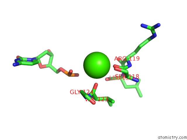
Mono view
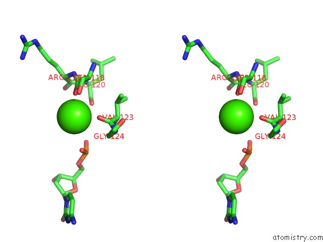
Stereo pair view
Mono view
Stereo pair view
A full contact list of Calcium with other atoms in the Ca binding
site number 1 of Crystal Structure of Muty Bound to Its Inhibitor Dna within 5.0Å range:
|
Reference:
S.Lee,
G.L.Verdine.
Atomic Substitution Reveals the Structural Basis For Substrate Adenine Recognition and Removal By Adenine Dna Glycosylase. Proc.Natl.Acad.Sci.Usa V. 106 18497 2009.
ISSN: ISSN 0027-8424
PubMed: 19841264
DOI: 10.1073/PNAS.0902908106
Page generated: Sat Jul 13 10:27:49 2024
ISSN: ISSN 0027-8424
PubMed: 19841264
DOI: 10.1073/PNAS.0902908106
Last articles
Zn in 9J0NZn in 9J0O
Zn in 9J0P
Zn in 9FJX
Zn in 9EKB
Zn in 9C0F
Zn in 9CAH
Zn in 9CH0
Zn in 9CH3
Zn in 9CH1