Calcium »
PDB 3fyi-3ghg »
3ga5 »
Calcium in PDB 3ga5: X-Ray Structure of Glucose/Galactose Receptor From Salmonella Typhimurium in Complex with (2R)-Glyceryl-Beta- D-Galactopyranoside
Protein crystallography data
The structure of X-Ray Structure of Glucose/Galactose Receptor From Salmonella Typhimurium in Complex with (2R)-Glyceryl-Beta- D-Galactopyranoside, PDB code: 3ga5
was solved by
S.Sooriyaarachchi,
W.Ubhayasekera,
S.L.Mowbray,
with X-Ray Crystallography technique. A brief refinement statistics is given in the table below:
| Resolution Low / High (Å) | 29.74 / 1.87 |
| Space group | P 21 21 21 |
| Cell size a, b, c (Å), α, β, γ (°) | 36.458, 109.275, 150.703, 90.00, 90.00, 90.00 |
| R / Rfree (%) | 17 / 22.2 |
Other elements in 3ga5:
The structure of X-Ray Structure of Glucose/Galactose Receptor From Salmonella Typhimurium in Complex with (2R)-Glyceryl-Beta- D-Galactopyranoside also contains other interesting chemical elements:
| Sodium | (Na) | 2 atoms |
Calcium Binding Sites:
The binding sites of Calcium atom in the X-Ray Structure of Glucose/Galactose Receptor From Salmonella Typhimurium in Complex with (2R)-Glyceryl-Beta- D-Galactopyranoside
(pdb code 3ga5). This binding sites where shown within
5.0 Angstroms radius around Calcium atom.
In total 2 binding sites of Calcium where determined in the X-Ray Structure of Glucose/Galactose Receptor From Salmonella Typhimurium in Complex with (2R)-Glyceryl-Beta- D-Galactopyranoside, PDB code: 3ga5:
Jump to Calcium binding site number: 1; 2;
In total 2 binding sites of Calcium where determined in the X-Ray Structure of Glucose/Galactose Receptor From Salmonella Typhimurium in Complex with (2R)-Glyceryl-Beta- D-Galactopyranoside, PDB code: 3ga5:
Jump to Calcium binding site number: 1; 2;
Calcium binding site 1 out of 2 in 3ga5
Go back to
Calcium binding site 1 out
of 2 in the X-Ray Structure of Glucose/Galactose Receptor From Salmonella Typhimurium in Complex with (2R)-Glyceryl-Beta- D-Galactopyranoside
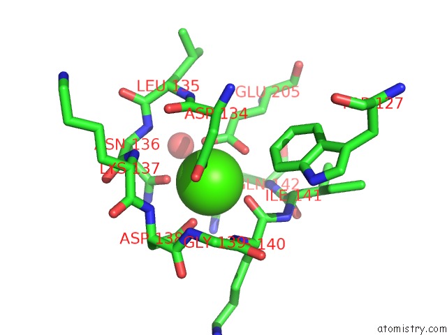
Mono view
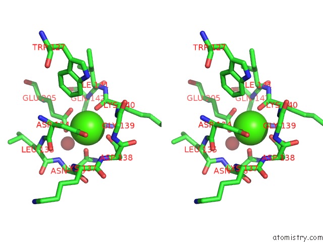
Stereo pair view
Mono view
Stereo pair view
A full contact list of Calcium with other atoms in the Ca binding
site number 1 of X-Ray Structure of Glucose/Galactose Receptor From Salmonella Typhimurium in Complex with (2R)-Glyceryl-Beta- D-Galactopyranoside within 5.0Å range:
|
Calcium binding site 2 out of 2 in 3ga5
Go back to
Calcium binding site 2 out
of 2 in the X-Ray Structure of Glucose/Galactose Receptor From Salmonella Typhimurium in Complex with (2R)-Glyceryl-Beta- D-Galactopyranoside
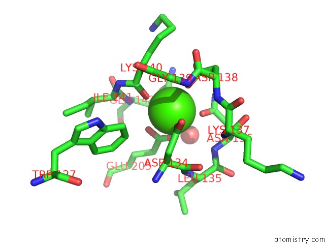
Mono view
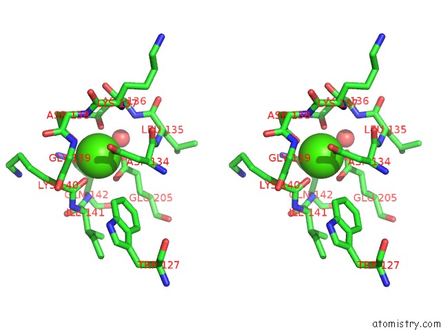
Stereo pair view
Mono view
Stereo pair view
A full contact list of Calcium with other atoms in the Ca binding
site number 2 of X-Ray Structure of Glucose/Galactose Receptor From Salmonella Typhimurium in Complex with (2R)-Glyceryl-Beta- D-Galactopyranoside within 5.0Å range:
|
Reference:
S.Sooriyaarachchi,
W.Ubhayasekera,
W.Boos,
S.L.Mowbray.
X-Ray Structure of Glucose/Galactose Receptor From Salmonella Typhimurium in Complex with the Physiological Ligand, (2R)-Glyceryl-Beta-D-Galactopyranoside Febs J. V. 276 2116 2009.
ISSN: ISSN 1742-464X
PubMed: 19292879
DOI: 10.1111/J.1742-4658.2009.06945.X
Page generated: Sat Jul 13 10:31:44 2024
ISSN: ISSN 1742-464X
PubMed: 19292879
DOI: 10.1111/J.1742-4658.2009.06945.X
Last articles
Zn in 9J0NZn in 9J0O
Zn in 9J0P
Zn in 9FJX
Zn in 9EKB
Zn in 9C0F
Zn in 9CAH
Zn in 9CH0
Zn in 9CH3
Zn in 9CH1