Calcium »
PDB 3gif-3gy5 »
3gk2 »
Calcium in PDB 3gk2: X-Ray Structure of Bovine SBI279,Ca(2+)-S100B
Protein crystallography data
The structure of X-Ray Structure of Bovine SBI279,Ca(2+)-S100B, PDB code: 3gk2
was solved by
T.H.Charpentier,
D.J.Weber,
E.A.Toth,
with X-Ray Crystallography technique. A brief refinement statistics is given in the table below:
| Resolution Low / High (Å) | 45.60 / 1.98 |
| Space group | C 2 2 21 |
| Cell size a, b, c (Å), α, β, γ (°) | 34.224, 91.158, 58.649, 90.00, 90.00, 90.00 |
| R / Rfree (%) | 22.7 / 28.1 |
Other elements in 3gk2:
The structure of X-Ray Structure of Bovine SBI279,Ca(2+)-S100B also contains other interesting chemical elements:
| Arsenic | (As) | 1 atom |
Calcium Binding Sites:
The binding sites of Calcium atom in the X-Ray Structure of Bovine SBI279,Ca(2+)-S100B
(pdb code 3gk2). This binding sites where shown within
5.0 Angstroms radius around Calcium atom.
In total 2 binding sites of Calcium where determined in the X-Ray Structure of Bovine SBI279,Ca(2+)-S100B, PDB code: 3gk2:
Jump to Calcium binding site number: 1; 2;
In total 2 binding sites of Calcium where determined in the X-Ray Structure of Bovine SBI279,Ca(2+)-S100B, PDB code: 3gk2:
Jump to Calcium binding site number: 1; 2;
Calcium binding site 1 out of 2 in 3gk2
Go back to
Calcium binding site 1 out
of 2 in the X-Ray Structure of Bovine SBI279,Ca(2+)-S100B
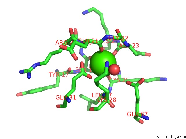
Mono view
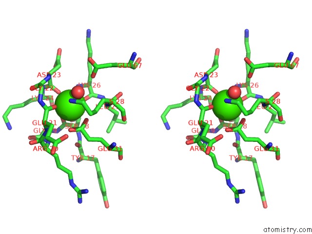
Stereo pair view
Mono view
Stereo pair view
A full contact list of Calcium with other atoms in the Ca binding
site number 1 of X-Ray Structure of Bovine SBI279,Ca(2+)-S100B within 5.0Å range:
|
Calcium binding site 2 out of 2 in 3gk2
Go back to
Calcium binding site 2 out
of 2 in the X-Ray Structure of Bovine SBI279,Ca(2+)-S100B
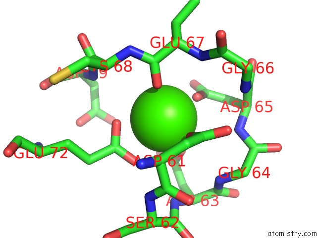
Mono view
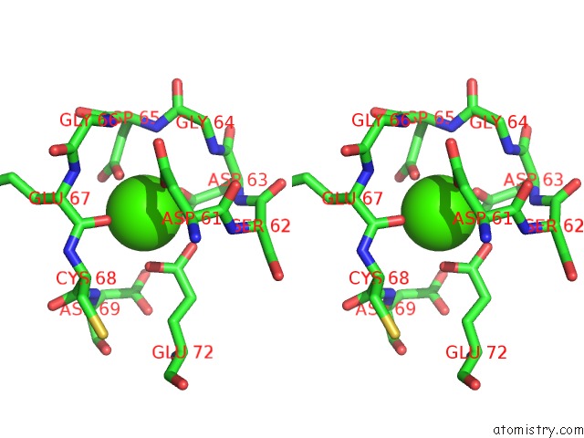
Stereo pair view
Mono view
Stereo pair view
A full contact list of Calcium with other atoms in the Ca binding
site number 2 of X-Ray Structure of Bovine SBI279,Ca(2+)-S100B within 5.0Å range:
|
Reference:
T.H.Charpentier,
P.T.Wilder,
M.A.Liriano,
K.M.Varney,
S.Zhong,
A.Coop,
E.Pozharski,
A.D.Mackerell,
E.A.Toth,
D.J.Weber.
Small Molecules Bound to Unique Sites in the Target Protein Binding Cleft of Calcium-Bound S100B As Characterized By Nuclear Magnetic Resonance and X-Ray Crystallography. Biochemistry V. 48 6202 2009.
ISSN: ISSN 0006-2960
PubMed: 19469484
DOI: 10.1021/BI9005754
Page generated: Sat Jul 13 10:42:46 2024
ISSN: ISSN 0006-2960
PubMed: 19469484
DOI: 10.1021/BI9005754
Last articles
Zn in 9J0NZn in 9J0O
Zn in 9J0P
Zn in 9FJX
Zn in 9EKB
Zn in 9C0F
Zn in 9CAH
Zn in 9CH0
Zn in 9CH3
Zn in 9CH1