Calcium »
PDB 3gif-3gy5 »
3gpe »
Calcium in PDB 3gpe: Crystal Structure Analysis of Pkc (Alpha)-C2 Domain Complexed with CA2+ and Ptdins(4,5)P2
Enzymatic activity of Crystal Structure Analysis of Pkc (Alpha)-C2 Domain Complexed with CA2+ and Ptdins(4,5)P2
All present enzymatic activity of Crystal Structure Analysis of Pkc (Alpha)-C2 Domain Complexed with CA2+ and Ptdins(4,5)P2:
2.7.11.13;
2.7.11.13;
Protein crystallography data
The structure of Crystal Structure Analysis of Pkc (Alpha)-C2 Domain Complexed with CA2+ and Ptdins(4,5)P2, PDB code: 3gpe
was solved by
C.Ferrer-Orta,
J.Querol-Audi,
I.Fita,
N.Verdaguer,
with X-Ray Crystallography technique. A brief refinement statistics is given in the table below:
| Resolution Low / High (Å) | 20.00 / 2.00 |
| Space group | P 32 2 1 |
| Cell size a, b, c (Å), α, β, γ (°) | 57.848, 57.848, 90.479, 90.00, 90.00, 120.00 |
| R / Rfree (%) | 24.3 / 27.7 |
Calcium Binding Sites:
The binding sites of Calcium atom in the Crystal Structure Analysis of Pkc (Alpha)-C2 Domain Complexed with CA2+ and Ptdins(4,5)P2
(pdb code 3gpe). This binding sites where shown within
5.0 Angstroms radius around Calcium atom.
In total 3 binding sites of Calcium where determined in the Crystal Structure Analysis of Pkc (Alpha)-C2 Domain Complexed with CA2+ and Ptdins(4,5)P2, PDB code: 3gpe:
Jump to Calcium binding site number: 1; 2; 3;
In total 3 binding sites of Calcium where determined in the Crystal Structure Analysis of Pkc (Alpha)-C2 Domain Complexed with CA2+ and Ptdins(4,5)P2, PDB code: 3gpe:
Jump to Calcium binding site number: 1; 2; 3;
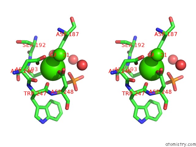
Calcium binding site 1 out of 3 in 3gpe
Go back to
Calcium binding site 1 out
of 3 in the Crystal Structure Analysis of Pkc (Alpha)-C2 Domain Complexed with CA2+ and Ptdins(4,5)P2

Mono view

Stereo pair view
Mono view
Stereo pair view
A full contact list of Calcium with other atoms in the Ca binding
site number 1 of Crystal Structure Analysis of Pkc (Alpha)-C2 Domain Complexed with CA2+ and Ptdins(4,5)P2 within 5.0Å range:
|
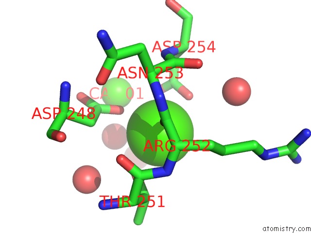
Calcium binding site 2 out of 3 in 3gpe
Go back to
Calcium binding site 2 out
of 3 in the Crystal Structure Analysis of Pkc (Alpha)-C2 Domain Complexed with CA2+ and Ptdins(4,5)P2

Mono view
Stereo pair view
Mono view
Stereo pair view
A full contact list of Calcium with other atoms in the Ca binding
site number 2 of Crystal Structure Analysis of Pkc (Alpha)-C2 Domain Complexed with CA2+ and Ptdins(4,5)P2 within 5.0Å range:
|
Calcium binding site 3 out of 3 in 3gpe
Go back to
Calcium binding site 3 out
of 3 in the Crystal Structure Analysis of Pkc (Alpha)-C2 Domain Complexed with CA2+ and Ptdins(4,5)P2
Mono view
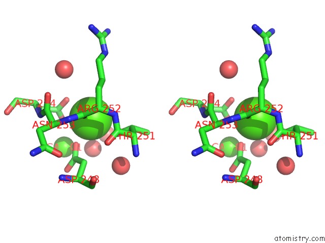
Stereo pair view
Mono view
Stereo pair view
A full contact list of Calcium with other atoms in the Ca binding
site number 3 of Crystal Structure Analysis of Pkc (Alpha)-C2 Domain Complexed with CA2+ and Ptdins(4,5)P2 within 5.0Å range:
|
Reference:
M.Guerrero-Valero,
C.Ferrer-Orta,
J.Querol-Audi,
C.Marin-Vicente,
I.Fita,
J.C.Gomez-Fernandez,
N.Verdaguer,
S.Corbalan-Garcia.
Structural and Mechanistic Insights Into the Association of Pkcalpha-C2 Domain to Ptdins(4,5)P2. Proc.Natl.Acad.Sci.Usa V. 106 6603 2009.
ISSN: ISSN 0027-8424
PubMed: 19346474
DOI: 10.1073/PNAS.0813099106
Page generated: Sat Jul 13 10:45:32 2024
ISSN: ISSN 0027-8424
PubMed: 19346474
DOI: 10.1073/PNAS.0813099106
Last articles
Zn in 9J0NZn in 9J0O
Zn in 9J0P
Zn in 9FJX
Zn in 9EKB
Zn in 9C0F
Zn in 9CAH
Zn in 9CH0
Zn in 9CH3
Zn in 9CH1