Calcium »
PDB 3gif-3gy5 »
3gwz »
Calcium in PDB 3gwz: Structure of the Mitomycin 7-O-Methyltransferase Mmcr
Protein crystallography data
The structure of Structure of the Mitomycin 7-O-Methyltransferase Mmcr, PDB code: 3gwz
was solved by
S.Singh,
A.Chang,
C.A.Bingman,
G.N.Phillips Jr.,
J.S.Thorson,
with X-Ray Crystallography technique. A brief refinement statistics is given in the table below:
| Resolution Low / High (Å) | 19.65 / 1.91 |
| Space group | P 21 21 21 |
| Cell size a, b, c (Å), α, β, γ (°) | 88.372, 98.918, 171.119, 90.00, 90.00, 90.00 |
| R / Rfree (%) | 17.4 / 21 |
Calcium Binding Sites:
The binding sites of Calcium atom in the Structure of the Mitomycin 7-O-Methyltransferase Mmcr
(pdb code 3gwz). This binding sites where shown within
5.0 Angstroms radius around Calcium atom.
In total 4 binding sites of Calcium where determined in the Structure of the Mitomycin 7-O-Methyltransferase Mmcr, PDB code: 3gwz:
Jump to Calcium binding site number: 1; 2; 3; 4;
In total 4 binding sites of Calcium where determined in the Structure of the Mitomycin 7-O-Methyltransferase Mmcr, PDB code: 3gwz:
Jump to Calcium binding site number: 1; 2; 3; 4;
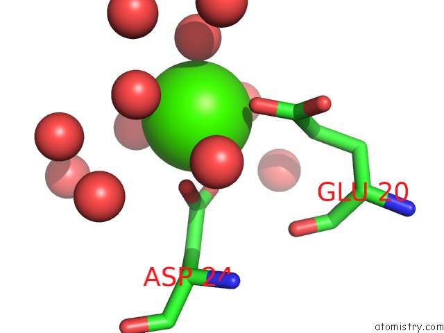
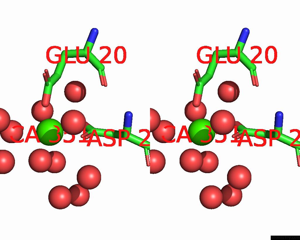
Calcium binding site 1 out of 4 in 3gwz
Go back to
Calcium binding site 1 out
of 4 in the Structure of the Mitomycin 7-O-Methyltransferase Mmcr
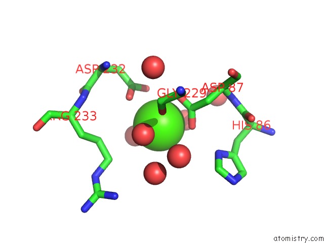
Mono view
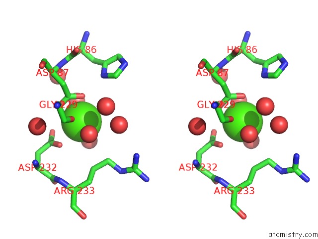
Stereo pair view
Mono view
Stereo pair view
A full contact list of Calcium with other atoms in the Ca binding
site number 1 of Structure of the Mitomycin 7-O-Methyltransferase Mmcr within 5.0Å range:
|
Calcium binding site 2 out of 4 in 3gwz
Go back to
Calcium binding site 2 out
of 4 in the Structure of the Mitomycin 7-O-Methyltransferase Mmcr
Mono view
Stereo pair view
Mono view
Stereo pair view
A full contact list of Calcium with other atoms in the Ca binding
site number 2 of Structure of the Mitomycin 7-O-Methyltransferase Mmcr within 5.0Å range:
|
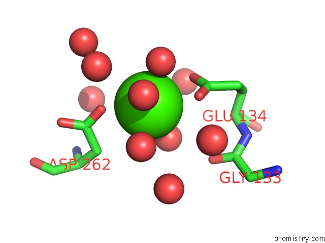
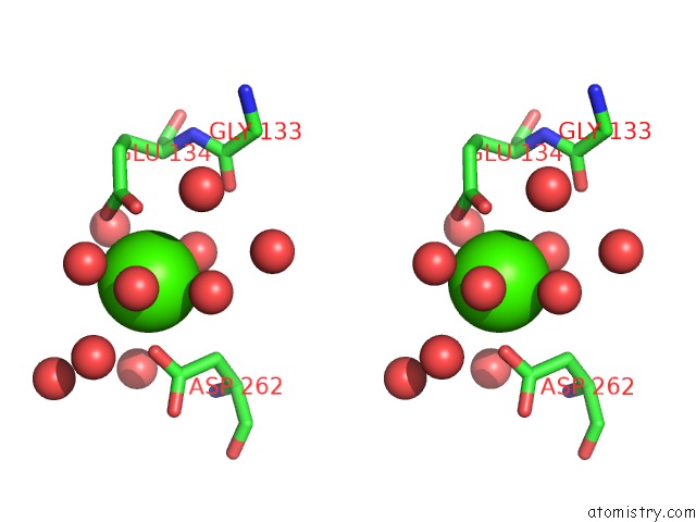
Calcium binding site 3 out of 4 in 3gwz
Go back to
Calcium binding site 3 out
of 4 in the Structure of the Mitomycin 7-O-Methyltransferase Mmcr
Mono view
Stereo pair view
Mono view
Stereo pair view
A full contact list of Calcium with other atoms in the Ca binding
site number 3 of Structure of the Mitomycin 7-O-Methyltransferase Mmcr within 5.0Å range:
|
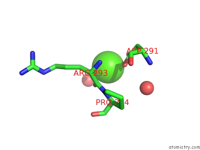
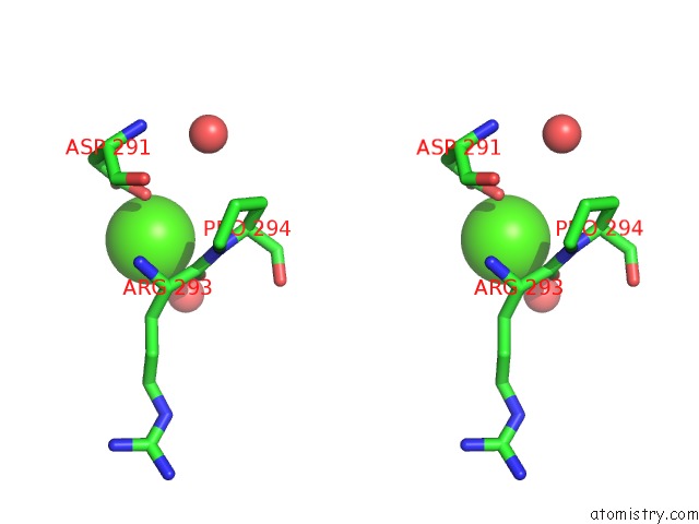
Calcium binding site 4 out of 4 in 3gwz
Go back to
Calcium binding site 4 out
of 4 in the Structure of the Mitomycin 7-O-Methyltransferase Mmcr
Mono view
Stereo pair view
Mono view
Stereo pair view
A full contact list of Calcium with other atoms in the Ca binding
site number 4 of Structure of the Mitomycin 7-O-Methyltransferase Mmcr within 5.0Å range:
|
Reference:
S.Singh,
A.Chang,
R.D.Goff,
C.A.Bingman,
S.Gruschow,
D.H.Sherman,
G.N.Phillips,
J.S.Thorson.
Structural Characterization of the Mitomycin 7-O-Methyltransferase. Proteins V. 79 2181 2011.
ISSN: ISSN 0887-3585
PubMed: 21538548
DOI: 10.1002/PROT.23040
Page generated: Sat Jul 13 10:49:00 2024
ISSN: ISSN 0887-3585
PubMed: 21538548
DOI: 10.1002/PROT.23040
Last articles
Zn in 9J0NZn in 9J0O
Zn in 9J0P
Zn in 9FJX
Zn in 9EKB
Zn in 9C0F
Zn in 9CAH
Zn in 9CH0
Zn in 9CH3
Zn in 9CH1