Calcium »
PDB 3gy6-3hii »
3gy6 »
Calcium in PDB 3gy6: A Comparative Study on the Inhibition of Bovine Beta-Trypsin By the Bis-Benzamidines Diminazene and Pentamidine
Enzymatic activity of A Comparative Study on the Inhibition of Bovine Beta-Trypsin By the Bis-Benzamidines Diminazene and Pentamidine
All present enzymatic activity of A Comparative Study on the Inhibition of Bovine Beta-Trypsin By the Bis-Benzamidines Diminazene and Pentamidine:
3.4.21.4;
3.4.21.4;
Protein crystallography data
The structure of A Comparative Study on the Inhibition of Bovine Beta-Trypsin By the Bis-Benzamidines Diminazene and Pentamidine, PDB code: 3gy6
was solved by
C.S.Perilo,
M.T.Pereira,
M.M.Santoro,
R.A.P.Nagem,
with X-Ray Crystallography technique. A brief refinement statistics is given in the table below:
| Resolution Low / High (Å) | 46.47 / 1.70 |
| Space group | P 31 2 1 |
| Cell size a, b, c (Å), α, β, γ (°) | 53.664, 53.664, 104.159, 90.00, 90.00, 120.00 |
| R / Rfree (%) | 17.7 / 22.7 |
Calcium Binding Sites:
The binding sites of Calcium atom in the A Comparative Study on the Inhibition of Bovine Beta-Trypsin By the Bis-Benzamidines Diminazene and Pentamidine
(pdb code 3gy6). This binding sites where shown within
5.0 Angstroms radius around Calcium atom.
In total only one binding site of Calcium was determined in the A Comparative Study on the Inhibition of Bovine Beta-Trypsin By the Bis-Benzamidines Diminazene and Pentamidine, PDB code: 3gy6:
In total only one binding site of Calcium was determined in the A Comparative Study on the Inhibition of Bovine Beta-Trypsin By the Bis-Benzamidines Diminazene and Pentamidine, PDB code: 3gy6:
Calcium binding site 1 out of 1 in 3gy6
Go back to
Calcium binding site 1 out
of 1 in the A Comparative Study on the Inhibition of Bovine Beta-Trypsin By the Bis-Benzamidines Diminazene and Pentamidine
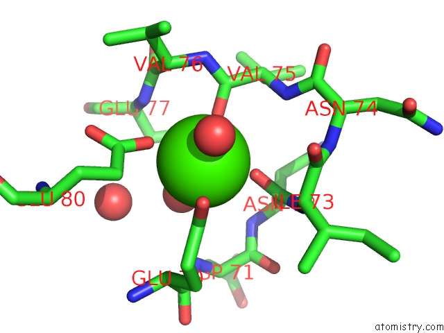
Mono view
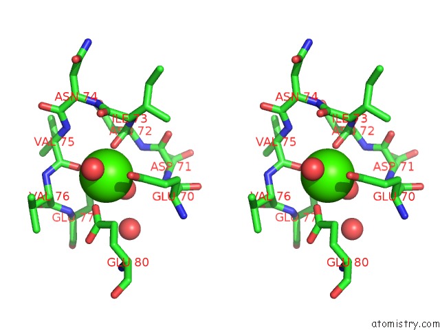
Stereo pair view
Mono view
Stereo pair view
A full contact list of Calcium with other atoms in the Ca binding
site number 1 of A Comparative Study on the Inhibition of Bovine Beta-Trypsin By the Bis-Benzamidines Diminazene and Pentamidine within 5.0Å range:
|
Reference:
C.S.Perilo,
M.T.Pereira,
M.M.Santoro,
R.A.Nagem.
Structural Binding Evidence of the Trypanocidal Drugs Berenil and Pentacarinate Active Principles to A Serine Protease Model. Int.J.Biol.Macromol. V. 46 502 2010.
ISSN: ISSN 0141-8130
PubMed: 20356563
DOI: 10.1016/J.IJBIOMAC.2010.03.006
Page generated: Sat Jul 13 10:51:35 2024
ISSN: ISSN 0141-8130
PubMed: 20356563
DOI: 10.1016/J.IJBIOMAC.2010.03.006
Last articles
Zn in 9J0NZn in 9J0O
Zn in 9J0P
Zn in 9FJX
Zn in 9EKB
Zn in 9C0F
Zn in 9CAH
Zn in 9CH0
Zn in 9CH3
Zn in 9CH1