Calcium »
PDB 3gy6-3hii »
3h3k »
Calcium in PDB 3h3k: Structure of A. Acidocaldarius Cellulase Cela in Complex with Cellotetraose
Enzymatic activity of Structure of A. Acidocaldarius Cellulase Cela in Complex with Cellotetraose
All present enzymatic activity of Structure of A. Acidocaldarius Cellulase Cela in Complex with Cellotetraose:
3.2.1.4;
3.2.1.4;
Protein crystallography data
The structure of Structure of A. Acidocaldarius Cellulase Cela in Complex with Cellotetraose, PDB code: 3h3k
was solved by
S.Morera,
K.Eckert,
with X-Ray Crystallography technique. A brief refinement statistics is given in the table below:
| Resolution Low / High (Å) | 30.22 / 2.10 |
| Space group | P 21 21 2 |
| Cell size a, b, c (Å), α, β, γ (°) | 85.090, 129.300, 49.130, 90.00, 90.00, 90.00 |
| R / Rfree (%) | 18.6 / 21.7 |
Other elements in 3h3k:
The structure of Structure of A. Acidocaldarius Cellulase Cela in Complex with Cellotetraose also contains other interesting chemical elements:
| Zinc | (Zn) | 1 atom |
Calcium Binding Sites:
The binding sites of Calcium atom in the Structure of A. Acidocaldarius Cellulase Cela in Complex with Cellotetraose
(pdb code 3h3k). This binding sites where shown within
5.0 Angstroms radius around Calcium atom.
In total only one binding site of Calcium was determined in the Structure of A. Acidocaldarius Cellulase Cela in Complex with Cellotetraose, PDB code: 3h3k:
In total only one binding site of Calcium was determined in the Structure of A. Acidocaldarius Cellulase Cela in Complex with Cellotetraose, PDB code: 3h3k:
Calcium binding site 1 out of 1 in 3h3k
Go back to
Calcium binding site 1 out
of 1 in the Structure of A. Acidocaldarius Cellulase Cela in Complex with Cellotetraose
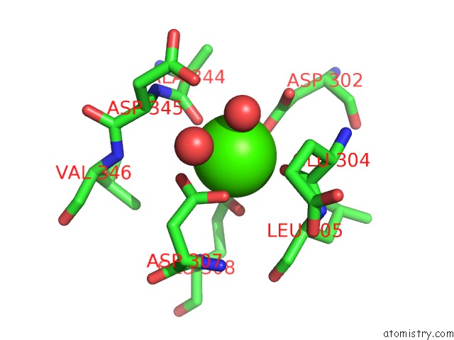
Mono view
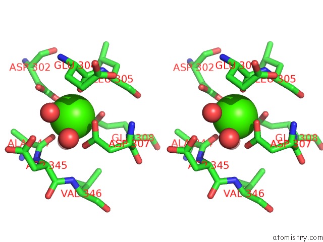
Stereo pair view
Mono view
Stereo pair view
A full contact list of Calcium with other atoms in the Ca binding
site number 1 of Structure of A. Acidocaldarius Cellulase Cela in Complex with Cellotetraose within 5.0Å range:
|
Reference:
K.Eckert,
A.Vigouroux,
L.Lo Leggio,
S.Morera.
Crystal Structures of A. Acidocaldarius Endoglucanase CEL9A in Complex with Cello-Oligosaccharides: Strong -1 and -2 Subsites Mimic Cellobiohydrolase Activity J.Mol.Biol. V. 394 61 2009.
ISSN: ISSN 0022-2836
PubMed: 19729024
DOI: 10.1016/J.JMB.2009.08.060
Page generated: Tue Jul 8 12:51:43 2025
ISSN: ISSN 0022-2836
PubMed: 19729024
DOI: 10.1016/J.JMB.2009.08.060
Last articles
Fe in 2YXOFe in 2YRS
Fe in 2YXC
Fe in 2YNM
Fe in 2YVJ
Fe in 2YP1
Fe in 2YU2
Fe in 2YU1
Fe in 2YQB
Fe in 2YOO