Calcium »
PDB 3gy6-3hii »
3haj »
Calcium in PDB 3haj: Crystal Structure of Human PACSIN2 F-Bar Domain (P212121 Lattice)
Protein crystallography data
The structure of Crystal Structure of Human PACSIN2 F-Bar Domain (P212121 Lattice), PDB code: 3haj
was solved by
Q.Wang,
M.V.A.S.Navarro,
G.Peng,
K.R.Rajashankar,
H.Sondermann,
with X-Ray Crystallography technique. A brief refinement statistics is given in the table below:
| Resolution Low / High (Å) | 44.72 / 2.78 |
| Space group | P 21 21 21 |
| Cell size a, b, c (Å), α, β, γ (°) | 31.295, 88.395, 357.722, 90.00, 90.00, 90.00 |
| R / Rfree (%) | 22.4 / 27.4 |
Calcium Binding Sites:
The binding sites of Calcium atom in the Crystal Structure of Human PACSIN2 F-Bar Domain (P212121 Lattice)
(pdb code 3haj). This binding sites where shown within
5.0 Angstroms radius around Calcium atom.
In total 2 binding sites of Calcium where determined in the Crystal Structure of Human PACSIN2 F-Bar Domain (P212121 Lattice), PDB code: 3haj:
Jump to Calcium binding site number: 1; 2;
In total 2 binding sites of Calcium where determined in the Crystal Structure of Human PACSIN2 F-Bar Domain (P212121 Lattice), PDB code: 3haj:
Jump to Calcium binding site number: 1; 2;
Calcium binding site 1 out of 2 in 3haj
Go back to
Calcium binding site 1 out
of 2 in the Crystal Structure of Human PACSIN2 F-Bar Domain (P212121 Lattice)
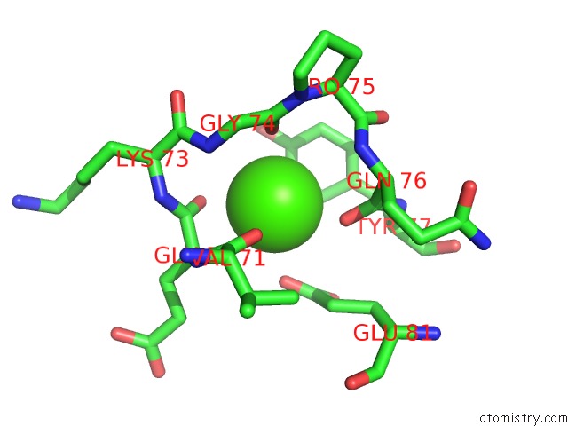
Mono view
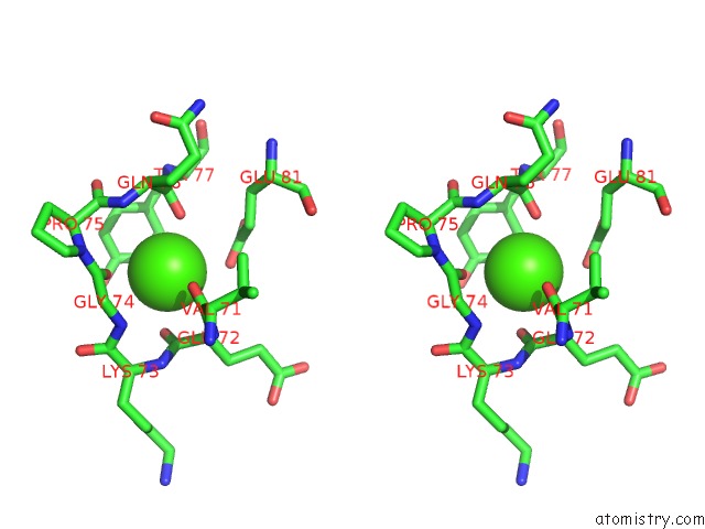
Stereo pair view
Mono view
Stereo pair view
A full contact list of Calcium with other atoms in the Ca binding
site number 1 of Crystal Structure of Human PACSIN2 F-Bar Domain (P212121 Lattice) within 5.0Å range:
|
Calcium binding site 2 out of 2 in 3haj
Go back to
Calcium binding site 2 out
of 2 in the Crystal Structure of Human PACSIN2 F-Bar Domain (P212121 Lattice)
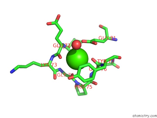
Mono view
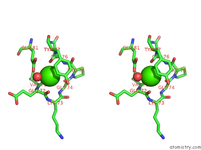
Stereo pair view
Mono view
Stereo pair view
A full contact list of Calcium with other atoms in the Ca binding
site number 2 of Crystal Structure of Human PACSIN2 F-Bar Domain (P212121 Lattice) within 5.0Å range:
|
Reference:
Q.Wang,
M.V.Navarro,
G.Peng,
E.Molinelli,
S.Lin Goh,
B.L.Judson,
K.R.Rajashankar,
H.Sondermann.
Molecular Mechanism of Membrane Constriction and Tubulation Mediated By the F-Bar Protein Pacsin/Syndapin. Proc.Natl.Acad.Sci.Usa V. 106 12700 2009.
ISSN: ISSN 0027-8424
PubMed: 19549836
DOI: 10.1073/PNAS.0902974106
Page generated: Sat Jul 13 10:54:03 2024
ISSN: ISSN 0027-8424
PubMed: 19549836
DOI: 10.1073/PNAS.0902974106
Last articles
Zn in 9J0NZn in 9J0O
Zn in 9J0P
Zn in 9FJX
Zn in 9EKB
Zn in 9C0F
Zn in 9CAH
Zn in 9CH0
Zn in 9CH3
Zn in 9CH1