Atomistry » Calcium » PDB 3gy6-3hii » 3hdb
Atomistry »
  Calcium »
    PDB 3gy6-3hii »
      3hdb »

Calcium in PDB 3hdb: Crystal Structure of Aahiv, A Metalloproteinase From Venom of Agkistrodon Acutus

Protein crystallography data

The structure of Crystal Structure of Aahiv, A Metalloproteinase From Venom of Agkistrodon Acutus, PDB code: 3hdb was solved by Z.Q.Zhu, L.W.Niu, M.K.Teng, with X-Ray Crystallography technique. A brief refinement statistics is given in the table below:

Resolution Low / High (Å) 38.27 / 2.31
Space group C 2 2 21
Cell size a, b, c (Å), α, β, γ (°) 111.190, 122.278, 94.721, 90.00, 90.00, 90.00
R / Rfree (%) 21.2 / 24.9

Other elements in 3hdb:

The structure of Crystal Structure of Aahiv, A Metalloproteinase From Venom of Agkistrodon Acutus also contains other interesting chemical elements:

Chlorine (Cl) 1 atom
Zinc (Zn) 1 atom

Calcium Binding Sites:

The binding sites of Calcium atom in the Crystal Structure of Aahiv, A Metalloproteinase From Venom of Agkistrodon Acutus (pdb code 3hdb). This binding sites where shown within 5.0 Angstroms radius around Calcium atom.
In total 7 binding sites of Calcium where determined in the Crystal Structure of Aahiv, A Metalloproteinase From Venom of Agkistrodon Acutus, PDB code: 3hdb:
Jump to Calcium binding site number: 1; 2; 3; 4; 5; 6; 7;

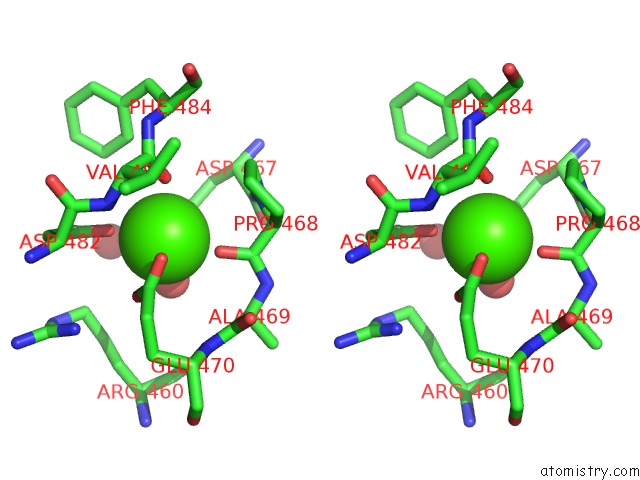
Calcium binding site 1 out of 7 in 3hdb

Go back to Calcium Binding Sites List in 3hdb
Calcium binding site 1 out of 7 in the Crystal Structure of Aahiv, A Metalloproteinase From Venom of Agkistrodon Acutus


Mono view


Stereo pair view

A full contact list of Calcium with other atoms in the Ca binding site number 1 of Crystal Structure of Aahiv, A Metalloproteinase From Venom of Agkistrodon Acutus within 5.0Å range:
probe atom residue distance (Å) B Occ
A:Ca621

b:38.3
occ:1.00
O A:HOH855 2.2 25.4 1.0
O A:CYS388 2.2 47.2 1.0
O A:HOH701 2.3 41.3 1.0
OE2 A:GLU200 2.4 35.6 1.0
OD1 A:ASN391 2.5 45.8 1.0
OD1 A:ASP284 2.5 38.4 1.0
OD2 A:ASP284 2.8 39.6 1.0
CG A:ASP284 3.0 38.4 1.0
CG A:ASN391 3.4 45.2 1.0
C A:CYS388 3.4 47.2 1.0
CD A:GLU200 3.5 35.5 1.0
ND2 A:ASN391 3.7 44.1 1.0
CG A:GLU200 4.0 35.3 1.0
O A:HOH844 4.3 44.0 1.0
CB A:GLU200 4.3 35.7 1.0
N A:ILE389 4.3 46.6 1.0
CA A:ILE389 4.3 46.4 1.0
O A:TYR198 4.4 41.0 1.0
CA A:CYS388 4.4 47.7 1.0
O A:HOH846 4.5 42.6 1.0
CB A:ASP284 4.5 38.2 1.0
OE1 A:GLU200 4.6 37.1 1.0
O A:ASN391 4.6 44.4 1.0
CB A:CYS388 4.6 47.5 1.0
CB A:ASN391 4.8 45.0 1.0
CE A:LYS282 4.9 50.0 1.0
N A:GLU200 4.9 36.6 1.0
C A:ILE389 4.9 46.3 1.0

Calcium binding site 2 out of 7 in 3hdb

Go back to Calcium Binding Sites List in 3hdb
Calcium binding site 2 out of 7 in the Crystal Structure of Aahiv, A Metalloproteinase From Venom of Agkistrodon Acutus


Mono view


Stereo pair view

A full contact list of Calcium with other atoms in the Ca binding site number 2 of Crystal Structure of Aahiv, A Metalloproteinase From Venom of Agkistrodon Acutus within 5.0Å range:
probe atom residue distance (Å) B Occ
A:Ca622

b:31.3
occ:1.00
OE2 A:GLU413 2.3 37.5 1.0
O A:LEU408 2.4 37.0 1.0
OD1 A:ASP416 2.4 36.8 1.0
OE2 A:GLU410 2.4 38.8 1.0
O A:LEU403 2.5 39.7 1.0
OD1 A:ASN406 2.5 40.6 1.0
OE1 A:GLU413 2.6 38.4 1.0
CD A:GLU413 2.8 38.2 1.0
CD A:GLU410 3.4 38.4 1.0
C A:LEU403 3.4 40.0 1.0
CG A:ASP416 3.4 36.7 1.0
CG A:ASN406 3.6 39.1 1.0
C A:LEU408 3.6 37.0 1.0
CG A:GLU410 4.0 37.9 1.0
CB A:ASP416 4.0 38.1 1.0
N A:LEU403 4.1 39.7 1.0
ND2 A:ASN406 4.1 38.1 1.0
CA A:CYS404 4.1 39.9 1.0
N A:LEU408 4.1 37.3 1.0
N A:CYS404 4.2 40.0 1.0
CB A:GLU410 4.2 37.0 1.0
CA A:LEU408 4.3 37.0 1.0
N A:GLY405 4.3 39.5 1.0
CG A:GLU413 4.3 37.8 1.0
N A:GLU410 4.3 36.8 1.0
CA A:LEU403 4.4 40.0 1.0
OE1 A:GLU410 4.4 37.4 1.0
N A:ASN406 4.4 38.9 1.0
OD2 A:ASP416 4.4 33.6 1.0
CB A:LEU408 4.5 36.9 1.0
N A:LEU409 4.6 36.7 1.0
C A:CYS404 4.7 39.9 1.0
CB A:ASN406 4.8 39.0 1.0
CA A:LEU409 4.8 36.7 1.0
O A:HOH830 4.9 32.2 1.0
N A:GLU407 4.9 38.5 1.0
C A:LEU409 4.9 36.6 1.0
CA A:GLU410 4.9 37.4 1.0
CA A:ASN406 5.0 38.9 1.0

Calcium binding site 3 out of 7 in 3hdb

Go back to Calcium Binding Sites List in 3hdb
Calcium binding site 3 out of 7 in the Crystal Structure of Aahiv, A Metalloproteinase From Venom of Agkistrodon Acutus


Mono view


Stereo pair view

A full contact list of Calcium with other atoms in the Ca binding site number 3 of Crystal Structure of Aahiv, A Metalloproteinase From Venom of Agkistrodon Acutus within 5.0Å range:
probe atom residue distance (Å) B Occ
A:Ca623

b:16.4
occ:1.00
OD1 A:ASP467 2.2 36.3 1.0
OD1 A:ASP482 2.2 36.1 1.0
O A:PRO468 2.3 32.9 1.0
O A:HOH742 2.5 27.5 1.0
OE2 A:GLU470 2.5 28.9 1.0
O A:VAL483 2.6 37.0 1.0
OE1 A:GLU470 2.6 28.5 1.0
CD A:GLU470 2.9 29.5 1.0
CG A:ASP467 3.3 34.2 1.0
CG A:ASP482 3.5 36.1 1.0
C A:PRO468 3.5 32.8 1.0
N A:VAL483 3.6 36.7 1.0
C A:VAL483 3.7 36.9 1.0
OD2 A:ASP467 3.7 34.4 1.0
OD2 A:ASP482 4.1 34.9 1.0
N A:PRO468 4.1 33.6 1.0
CA A:VAL483 4.2 36.8 1.0
C A:ASP467 4.3 33.8 1.0
CA A:PRO468 4.4 33.0 1.0
C A:ASP482 4.4 36.7 1.0
CA A:ASP482 4.4 36.4 1.0
N A:ALA469 4.4 32.4 1.0
CG A:GLU470 4.4 28.8 1.0
CG A:PRO468 4.4 33.6 1.0
CD A:PRO468 4.5 33.8 1.0
CA A:ALA469 4.5 31.8 1.0
CB A:ASP467 4.5 34.1 1.0
O A:HOH745 4.5 30.6 1.0
CB A:ASP482 4.5 36.2 1.0
C A:ALA469 4.6 31.3 1.0
CD1 A:PHE484 4.6 36.2 1.0
CA A:ASP467 4.6 34.0 1.0
O A:ASP467 4.7 33.5 1.0
CD A:ARG460 4.7 32.1 1.0
N A:GLU470 4.7 30.9 1.0
N A:PHE484 4.8 36.9 1.0
CE1 A:PHE484 4.8 36.5 1.0
CB A:VAL483 4.9 36.6 1.0
CB A:PRO468 4.9 33.0 1.0

Calcium binding site 4 out of 7 in 3hdb

Go back to Calcium Binding Sites List in 3hdb
Calcium binding site 4 out of 7 in the Crystal Structure of Aahiv, A Metalloproteinase From Venom of Agkistrodon Acutus


Mono view


Stereo pair view

A full contact list of Calcium with other atoms in the Ca binding site number 4 of Crystal Structure of Aahiv, A Metalloproteinase From Venom of Agkistrodon Acutus within 5.0Å range:
probe atom residue distance (Å) B Occ
A:Ca654

b:78.5
occ:1.00
O A:ALA279 3.8 51.6 1.0
O A:LEU278 3.9 50.6 1.0
C A:ALA279 4.1 51.4 1.0
N A:LYS281 4.2 49.8 1.0
C A:GLU280 4.4 50.5 1.0
N A:GLU280 4.6 51.4 1.0
N A:LYS282 4.6 46.8 1.0
C A:LYS281 4.6 48.0 1.0
CA A:ALA279 4.7 51.1 1.0
CA A:LYS281 4.8 49.0 1.0
CA A:GLU280 4.8 51.4 1.0
O A:GLU280 4.8 50.5 1.0
C A:LEU278 4.9 50.6 1.0

Calcium binding site 5 out of 7 in 3hdb

Go back to Calcium Binding Sites List in 3hdb
Calcium binding site 5 out of 7 in the Crystal Structure of Aahiv, A Metalloproteinase From Venom of Agkistrodon Acutus


Mono view


Stereo pair view

A full contact list of Calcium with other atoms in the Ca binding site number 5 of Crystal Structure of Aahiv, A Metalloproteinase From Venom of Agkistrodon Acutus within 5.0Å range:
probe atom residue distance (Å) B Occ
A:Ca655

b:79.5
occ:1.00
N A:ASP523 3.0 40.2 1.0
OE1 A:GLU522 3.3 49.6 1.0
CG A:ASP523 3.4 41.2 1.0
OD1 A:ASP523 3.6 44.7 1.0
CB A:ASP523 3.6 40.0 1.0
OD2 A:ASP523 3.6 40.5 1.0
CA A:GLU522 3.7 41.9 1.0
O A:HOH939 3.7 38.4 1.0
C A:GLU522 3.8 41.0 1.0
CA A:ASP523 3.9 39.4 1.0
CB A:GLU522 3.9 42.0 1.0
CD A:GLU522 4.1 48.3 1.0
CG A:GLU522 4.6 45.0 1.0
OE2 A:GLU522 4.9 49.5 1.0
N A:GLU522 5.0 42.1 1.0

Calcium binding site 6 out of 7 in 3hdb

Go back to Calcium Binding Sites List in 3hdb
Calcium binding site 6 out of 7 in the Crystal Structure of Aahiv, A Metalloproteinase From Venom of Agkistrodon Acutus


Mono view


Stereo pair view

A full contact list of Calcium with other atoms in the Ca binding site number 6 of Crystal Structure of Aahiv, A Metalloproteinase From Venom of Agkistrodon Acutus within 5.0Å range:
probe atom residue distance (Å) B Occ
A:Ca656

b:49.1
occ:1.00
O A:LEU558 2.7 26.4 1.0
N A:PHE574 2.8 28.1 1.0
O A:GLY583 2.8 27.2 1.0
CA A:MET573 3.4 28.7 1.0
CB A:MET573 3.5 28.8 1.0
CA A:GLY583 3.5 27.7 1.0
C A:MET573 3.5 28.6 1.0
C A:GLY583 3.5 27.3 1.0
C A:LEU558 3.6 26.7 1.0
CD A:ARG557 3.6 23.1 1.0
O A:PHE574 3.7 28.6 1.0
N A:LEU558 3.7 26.9 1.0
CA A:PHE574 3.8 27.8 1.0
CB A:LEU558 3.8 26.6 1.0
CA A:LEU558 3.9 26.6 1.0
CG A:ARG557 3.9 25.1 1.0
CB A:PHE574 4.0 27.7 1.0
CD1 A:LEU513 4.1 36.7 1.0
C A:PHE574 4.2 28.1 1.0
CG A:LEU558 4.5 26.5 1.0
CZ A:PHE514 4.7 36.3 1.0
O A:MET573 4.7 28.3 1.0
C A:ARG557 4.7 26.5 1.0
N A:MET573 4.8 29.8 1.0
N A:TYR559 4.8 27.6 1.0
CD2 A:LEU558 4.8 25.7 1.0
N A:MET584 4.8 26.8 1.0
CE1 A:PHE514 4.8 36.6 1.0
CG A:MET573 4.8 27.3 1.0
O A:LYS572 4.9 30.4 1.0
N A:GLY583 4.9 28.2 1.0
NE A:ARG557 5.0 23.5 1.0

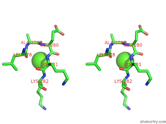
Calcium binding site 7 out of 7 in 3hdb

Go back to Calcium Binding Sites List in 3hdb
Calcium binding site 7 out of 7 in the Crystal Structure of Aahiv, A Metalloproteinase From Venom of Agkistrodon Acutus


Mono view


Stereo pair view

A full contact list of Calcium with other atoms in the Ca binding site number 7 of Crystal Structure of Aahiv, A Metalloproteinase From Venom of Agkistrodon Acutus within 5.0Å range:
probe atom residue distance (Å) B Occ
A:Ca657

b:73.4
occ:1.00
O A:HOH831 2.5 34.8 1.0
O A:PHE294 2.5 38.7 1.0
O A:GLY296 2.6 43.6 1.0
OD1 A:ASP293 2.7 41.9 1.0
NZ L:LYS1 3.3 59.2 1.0
CG A:ASP293 3.3 38.7 1.0
O A:HOH836 3.3 44.4 1.0
OD2 A:ASP293 3.5 40.2 1.0
C A:PHE294 3.6 38.9 1.0
C A:GLY296 3.8 43.6 1.0
N A:PHE294 4.0 37.3 1.0
N A:GLY296 4.2 42.8 1.0
C A:ASP293 4.3 36.7 1.0
OE1 A:GLN318 4.3 37.5 1.0
CA A:PHE294 4.4 38.0 1.0
C A:ARG295 4.4 42.2 1.0
CB A:ASP293 4.5 36.8 1.0
O A:ILE298 4.6 42.1 1.0
CA A:GLY296 4.6 43.4 1.0
N A:ARG295 4.6 40.3 1.0
CA A:ASP293 4.6 36.4 1.0
CE L:LYS1 4.7 59.3 1.0
N A:SER297 4.7 44.0 1.0
O A:ASP293 4.8 37.1 1.0
NE2 A:GLN318 4.8 39.1 1.0
CA A:ARG295 4.8 41.9 1.0
O A:ARG295 4.8 42.4 1.0
CB A:PHE294 4.8 37.7 1.0
CA A:SER297 4.8 44.5 1.0

Reference:

Z.Zhu, Y.Gao, Z.Zhu, Y.Yu, X.Zhang, J.Zang, M.Teng, L.Niu. Structural Basis of the Autolysis of Aahiv Suggests A Novel Target Recognizing Model For Adam/Reprolysin Family Proteins Biochem.Biophys.Res.Commun. V. 386 159 2009.
ISSN: ISSN 0006-291X
PubMed: 19505434
DOI: 10.1016/J.BBRC.2009.06.004
Page generated: Sat Dec 12 04:13:56 2020

Last articles

Zn in 8WB0
Zn in 8WAX
Zn in 8WAU
Zn in 8WAZ
Zn in 8WAY
Zn in 8WAV
Zn in 8WAW
Zn in 8WAT
Zn in 8W7M
Zn in 8WD3
© Copyright 2008-2020 by atomistry.com
Home   |    Site Map   |    Copyright   |    Contact us   |    Privacy