Calcium »
PDB 3hjb-3hz3 »
3hus »
Calcium in PDB 3hus: Crystal Structure of Recombinant Gamma N308K Fibrinogen Fragment D with the Peptide Ligand Gly-Pro-Arg-Pro-Amide
Protein crystallography data
The structure of Crystal Structure of Recombinant Gamma N308K Fibrinogen Fragment D with the Peptide Ligand Gly-Pro-Arg-Pro-Amide, PDB code: 3hus
was solved by
S.T.Lord,
S.R.Bowley,
N.Okumura,
with X-Ray Crystallography technique. A brief refinement statistics is given in the table below:
| Resolution Low / High (Å) | 47.25 / 3.04 |
| Space group | P 43 21 2 |
| Cell size a, b, c (Å), α, β, γ (°) | 95.009, 95.009, 448.382, 90.00, 90.00, 90.00 |
| R / Rfree (%) | 22 / 28.8 |
Calcium Binding Sites:
The binding sites of Calcium atom in the Crystal Structure of Recombinant Gamma N308K Fibrinogen Fragment D with the Peptide Ligand Gly-Pro-Arg-Pro-Amide
(pdb code 3hus). This binding sites where shown within
5.0 Angstroms radius around Calcium atom.
In total 4 binding sites of Calcium where determined in the Crystal Structure of Recombinant Gamma N308K Fibrinogen Fragment D with the Peptide Ligand Gly-Pro-Arg-Pro-Amide, PDB code: 3hus:
Jump to Calcium binding site number: 1; 2; 3; 4;
In total 4 binding sites of Calcium where determined in the Crystal Structure of Recombinant Gamma N308K Fibrinogen Fragment D with the Peptide Ligand Gly-Pro-Arg-Pro-Amide, PDB code: 3hus:
Jump to Calcium binding site number: 1; 2; 3; 4;
Calcium binding site 1 out of 4 in 3hus
Go back to
Calcium binding site 1 out
of 4 in the Crystal Structure of Recombinant Gamma N308K Fibrinogen Fragment D with the Peptide Ligand Gly-Pro-Arg-Pro-Amide

Mono view
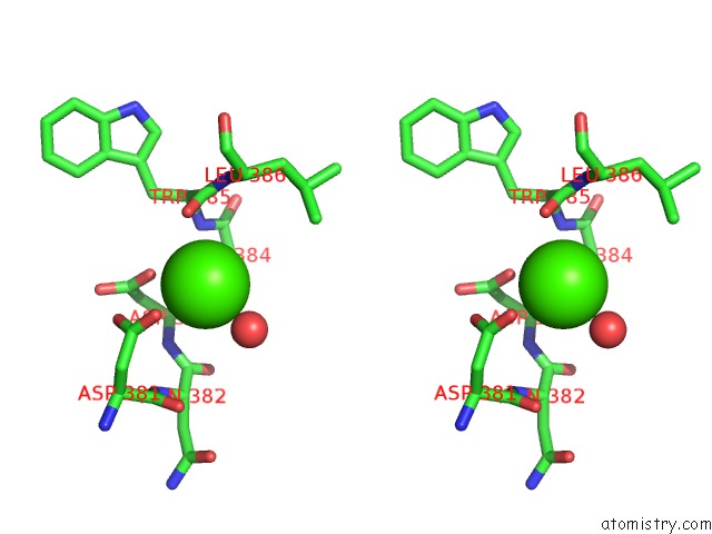
Stereo pair view
Mono view
Stereo pair view
A full contact list of Calcium with other atoms in the Ca binding
site number 1 of Crystal Structure of Recombinant Gamma N308K Fibrinogen Fragment D with the Peptide Ligand Gly-Pro-Arg-Pro-Amide within 5.0Å range:
|
Calcium binding site 2 out of 4 in 3hus
Go back to
Calcium binding site 2 out
of 4 in the Crystal Structure of Recombinant Gamma N308K Fibrinogen Fragment D with the Peptide Ligand Gly-Pro-Arg-Pro-Amide
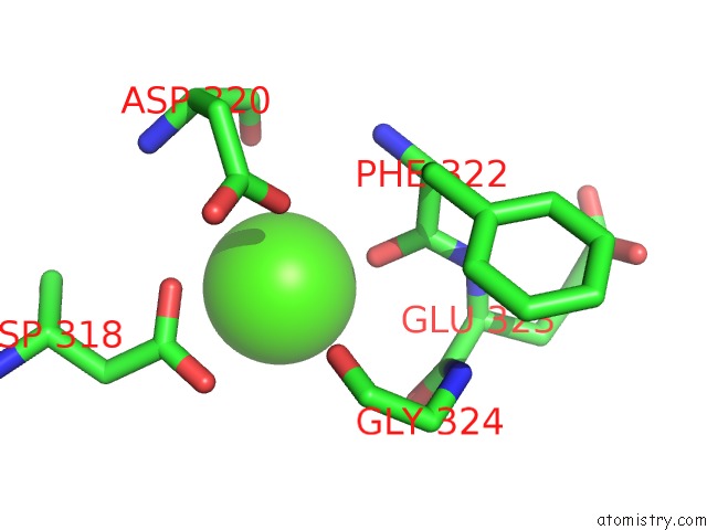
Mono view

Stereo pair view
Mono view
Stereo pair view
A full contact list of Calcium with other atoms in the Ca binding
site number 2 of Crystal Structure of Recombinant Gamma N308K Fibrinogen Fragment D with the Peptide Ligand Gly-Pro-Arg-Pro-Amide within 5.0Å range:
|
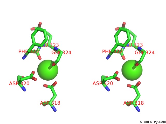
Calcium binding site 3 out of 4 in 3hus
Go back to
Calcium binding site 3 out
of 4 in the Crystal Structure of Recombinant Gamma N308K Fibrinogen Fragment D with the Peptide Ligand Gly-Pro-Arg-Pro-Amide
Mono view

Stereo pair view
Mono view
Stereo pair view
A full contact list of Calcium with other atoms in the Ca binding
site number 3 of Crystal Structure of Recombinant Gamma N308K Fibrinogen Fragment D with the Peptide Ligand Gly-Pro-Arg-Pro-Amide within 5.0Å range:
|
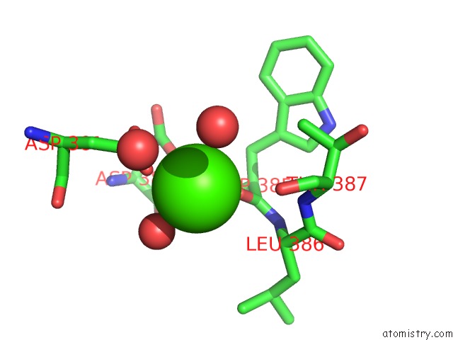
Calcium binding site 4 out of 4 in 3hus
Go back to
Calcium binding site 4 out
of 4 in the Crystal Structure of Recombinant Gamma N308K Fibrinogen Fragment D with the Peptide Ligand Gly-Pro-Arg-Pro-Amide

Mono view
Stereo pair view
Mono view
Stereo pair view
A full contact list of Calcium with other atoms in the Ca binding
site number 4 of Crystal Structure of Recombinant Gamma N308K Fibrinogen Fragment D with the Peptide Ligand Gly-Pro-Arg-Pro-Amide within 5.0Å range:
|
Reference:
S.R.Bowley,
N.Okumura,
S.T.Lord.
Impaired Protofibril Formation in Fibrinogen GAMMAN308K Is Due to Altered D:D and "A:A" Interactions. Biochemistry V. 48 8656 2009.
ISSN: ISSN 0006-2960
PubMed: 19650644
DOI: 10.1021/BI900239B
Page generated: Sat Jul 13 11:12:35 2024
ISSN: ISSN 0006-2960
PubMed: 19650644
DOI: 10.1021/BI900239B
Last articles
Zn in 9MJ5Zn in 9HNW
Zn in 9G0L
Zn in 9FNE
Zn in 9DZN
Zn in 9E0I
Zn in 9D32
Zn in 9DAK
Zn in 8ZXC
Zn in 8ZUF