Calcium »
PDB 3hjb-3hz3 »
3hx4 »
Calcium in PDB 3hx4: Crystal Structure of CDPK1 of Toxoplasma Gondii, TGME49_101440, in Presence of Calcium
Protein crystallography data
The structure of Crystal Structure of CDPK1 of Toxoplasma Gondii, TGME49_101440, in Presence of Calcium, PDB code: 3hx4
was solved by
A.K.Wernimont,
J.D.Artz,
P.Finnerty,
T.Xiao,
H.He,
F.Mackenzie,
G.Sinestera,
A.A.Hassani,
G.Wasney,
M.Vedadi,
S.Lourido,
A.Bochkarev,
C.H.Arrowsmith,
A.M.Edwards,
C.Bountra,
J.Weigelt,
D.L.Sibley,
R.Hui,
Y.H.Lin,
Structural Genomics Consortium (Sgc),
with X-Ray Crystallography technique. A brief refinement statistics is given in the table below:
| Resolution Low / High (Å) | 50.00 / 1.95 |
| Space group | P 21 21 21 |
| Cell size a, b, c (Å), α, β, γ (°) | 49.236, 95.605, 101.612, 90.00, 90.00, 90.00 |
| R / Rfree (%) | 20.9 / 25.7 |
Calcium Binding Sites:
The binding sites of Calcium atom in the Crystal Structure of CDPK1 of Toxoplasma Gondii, TGME49_101440, in Presence of Calcium
(pdb code 3hx4). This binding sites where shown within
5.0 Angstroms radius around Calcium atom.
In total 4 binding sites of Calcium where determined in the Crystal Structure of CDPK1 of Toxoplasma Gondii, TGME49_101440, in Presence of Calcium, PDB code: 3hx4:
Jump to Calcium binding site number: 1; 2; 3; 4;
In total 4 binding sites of Calcium where determined in the Crystal Structure of CDPK1 of Toxoplasma Gondii, TGME49_101440, in Presence of Calcium, PDB code: 3hx4:
Jump to Calcium binding site number: 1; 2; 3; 4;
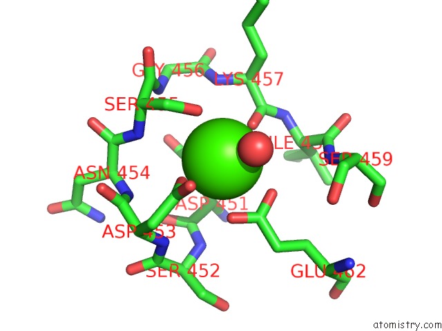
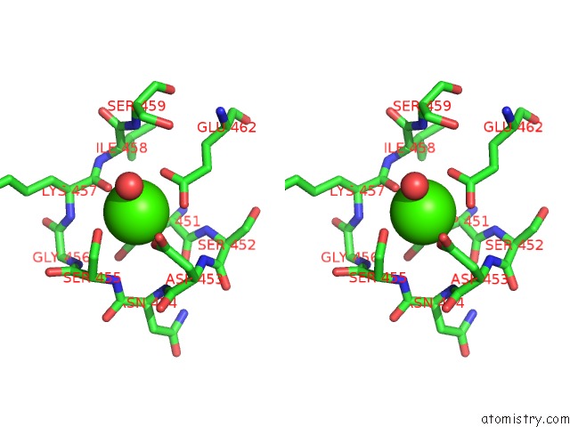
Calcium binding site 1 out of 4 in 3hx4
Go back to
Calcium binding site 1 out
of 4 in the Crystal Structure of CDPK1 of Toxoplasma Gondii, TGME49_101440, in Presence of Calcium
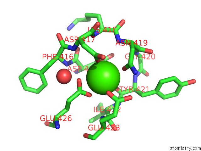
Mono view
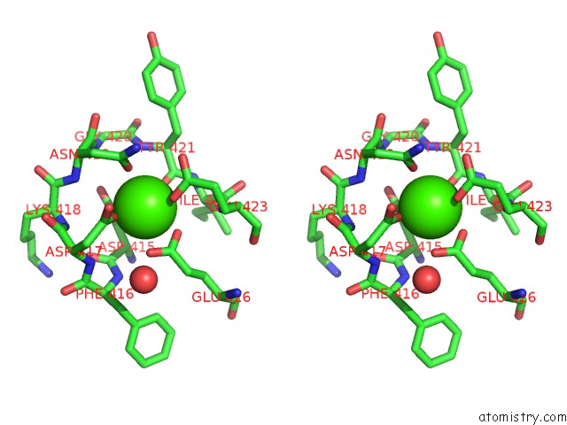
Stereo pair view
Mono view
Stereo pair view
A full contact list of Calcium with other atoms in the Ca binding
site number 1 of Crystal Structure of CDPK1 of Toxoplasma Gondii, TGME49_101440, in Presence of Calcium within 5.0Å range:
|
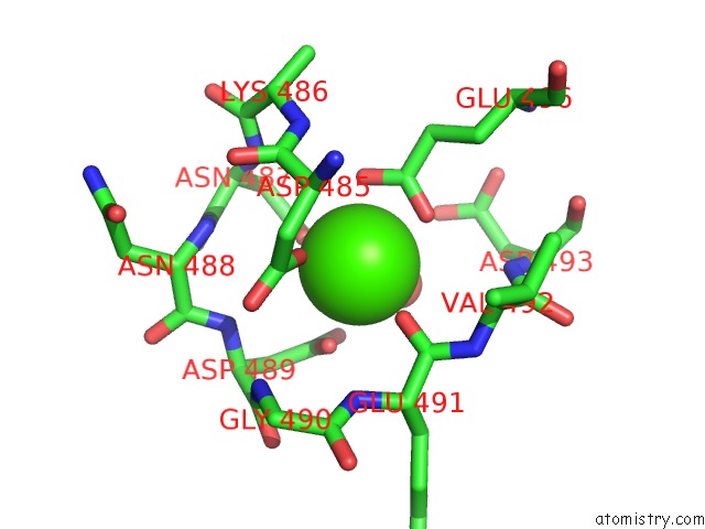
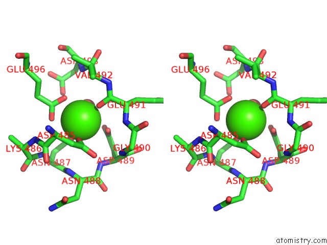
Calcium binding site 2 out of 4 in 3hx4
Go back to
Calcium binding site 2 out
of 4 in the Crystal Structure of CDPK1 of Toxoplasma Gondii, TGME49_101440, in Presence of Calcium
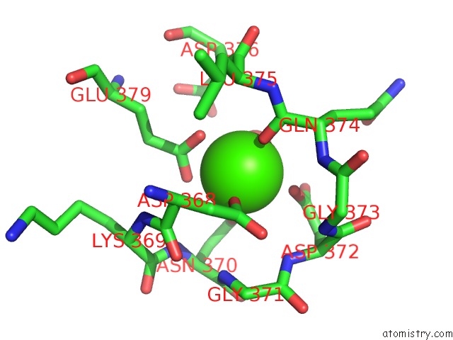
Mono view
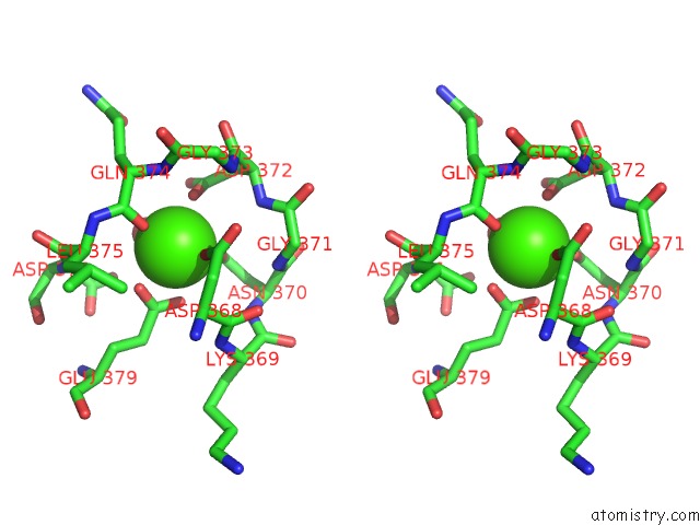
Stereo pair view
Mono view
Stereo pair view
A full contact list of Calcium with other atoms in the Ca binding
site number 2 of Crystal Structure of CDPK1 of Toxoplasma Gondii, TGME49_101440, in Presence of Calcium within 5.0Å range:
|
Calcium binding site 3 out of 4 in 3hx4
Go back to
Calcium binding site 3 out
of 4 in the Crystal Structure of CDPK1 of Toxoplasma Gondii, TGME49_101440, in Presence of Calcium
Mono view
Stereo pair view
Mono view
Stereo pair view
A full contact list of Calcium with other atoms in the Ca binding
site number 3 of Crystal Structure of CDPK1 of Toxoplasma Gondii, TGME49_101440, in Presence of Calcium within 5.0Å range:
|
Calcium binding site 4 out of 4 in 3hx4
Go back to
Calcium binding site 4 out
of 4 in the Crystal Structure of CDPK1 of Toxoplasma Gondii, TGME49_101440, in Presence of Calcium
Mono view
Stereo pair view
Mono view
Stereo pair view
A full contact list of Calcium with other atoms in the Ca binding
site number 4 of Crystal Structure of CDPK1 of Toxoplasma Gondii, TGME49_101440, in Presence of Calcium within 5.0Å range:
|
Reference:
A.K.Wernimont,
J.D.Artz,
P.Finerty,
Y.H.Lin,
M.Amani,
A.Allali-Hassani,
G.Senisterra,
M.Vedadi,
W.Tempel,
F.Mackenzie,
I.Chau,
S.Lourido,
L.D.Sibley,
R.Hui.
Structures of Apicomplexan Calcium-Dependent Protein Kinases Reveal Mechanism of Activation By Calcium. Nat.Struct.Mol.Biol. V. 17 596 2010.
ISSN: ISSN 1545-9993
PubMed: 20436473
DOI: 10.1038/NSMB.1795
Page generated: Sat Jul 13 11:12:52 2024
ISSN: ISSN 1545-9993
PubMed: 20436473
DOI: 10.1038/NSMB.1795
Last articles
Zn in 9MJ5Zn in 9HNW
Zn in 9G0L
Zn in 9FNE
Zn in 9DZN
Zn in 9E0I
Zn in 9D32
Zn in 9DAK
Zn in 8ZXC
Zn in 8ZUF