Calcium »
PDB 3hzb-3iit »
3icb »
Calcium in PDB 3icb: The Refined Structure of Vitamin D-Dependent Calcium- Binding Protein From Bovine Intestine. Molecular Details, Ion Binding, and Implications For the Structure of Other Calcium-Binding Proteins
Protein crystallography data
The structure of The Refined Structure of Vitamin D-Dependent Calcium- Binding Protein From Bovine Intestine. Molecular Details, Ion Binding, and Implications For the Structure of Other Calcium-Binding Proteins, PDB code: 3icb
was solved by
D.M.E.Szebenyi,
K.Moffat,
with X-Ray Crystallography technique. A brief refinement statistics is given in the table below:
| Resolution Low / High (Å) | 9.00 / 2.30 |
| Space group | P 21 21 2 |
| Cell size a, b, c (Å), α, β, γ (°) | 56.200, 42.600, 29.200, 90.00, 90.00, 90.00 |
| R / Rfree (%) | n/a / n/a |
Calcium Binding Sites:
The binding sites of Calcium atom in the The Refined Structure of Vitamin D-Dependent Calcium- Binding Protein From Bovine Intestine. Molecular Details, Ion Binding, and Implications For the Structure of Other Calcium-Binding Proteins
(pdb code 3icb). This binding sites where shown within
5.0 Angstroms radius around Calcium atom.
In total 2 binding sites of Calcium where determined in the The Refined Structure of Vitamin D-Dependent Calcium- Binding Protein From Bovine Intestine. Molecular Details, Ion Binding, and Implications For the Structure of Other Calcium-Binding Proteins, PDB code: 3icb:
Jump to Calcium binding site number: 1; 2;
Calcium binding site 1 out of 2 in 3icb
Go back to
Calcium binding site 1 out
of 2 in the The Refined Structure of Vitamin D-Dependent Calcium- Binding Protein From Bovine Intestine. Molecular Details, Ion Binding, and Implications For the Structure of Other Calcium-Binding Proteins
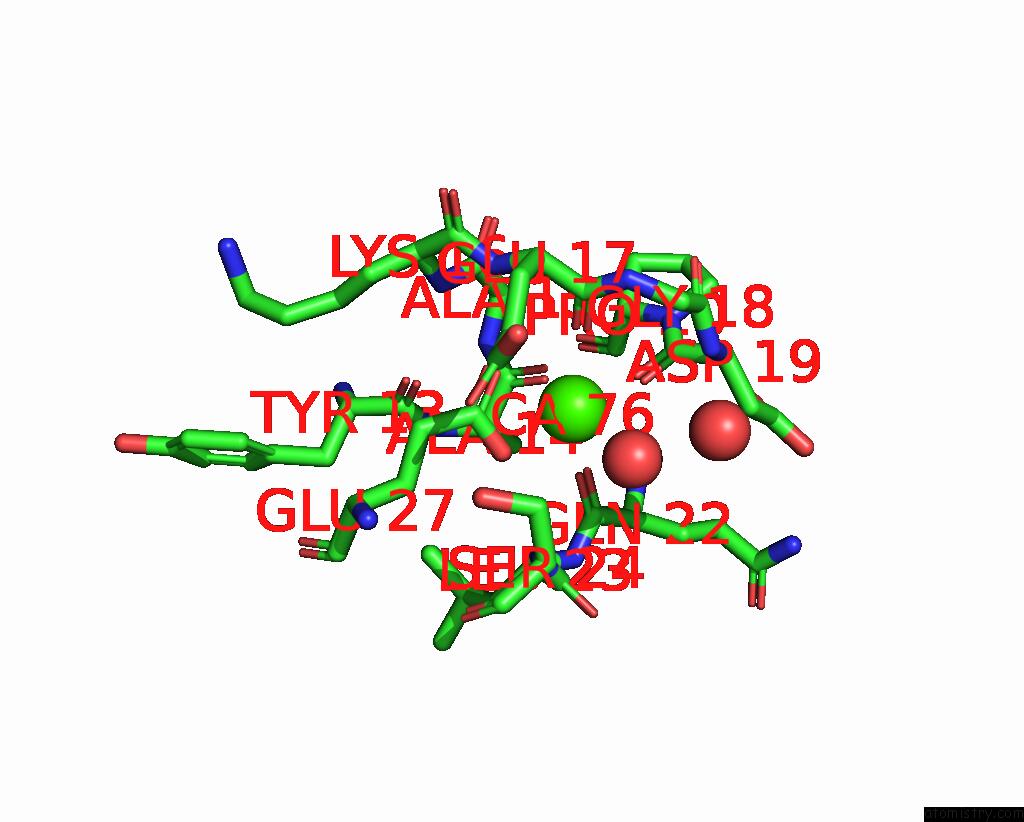
Mono view
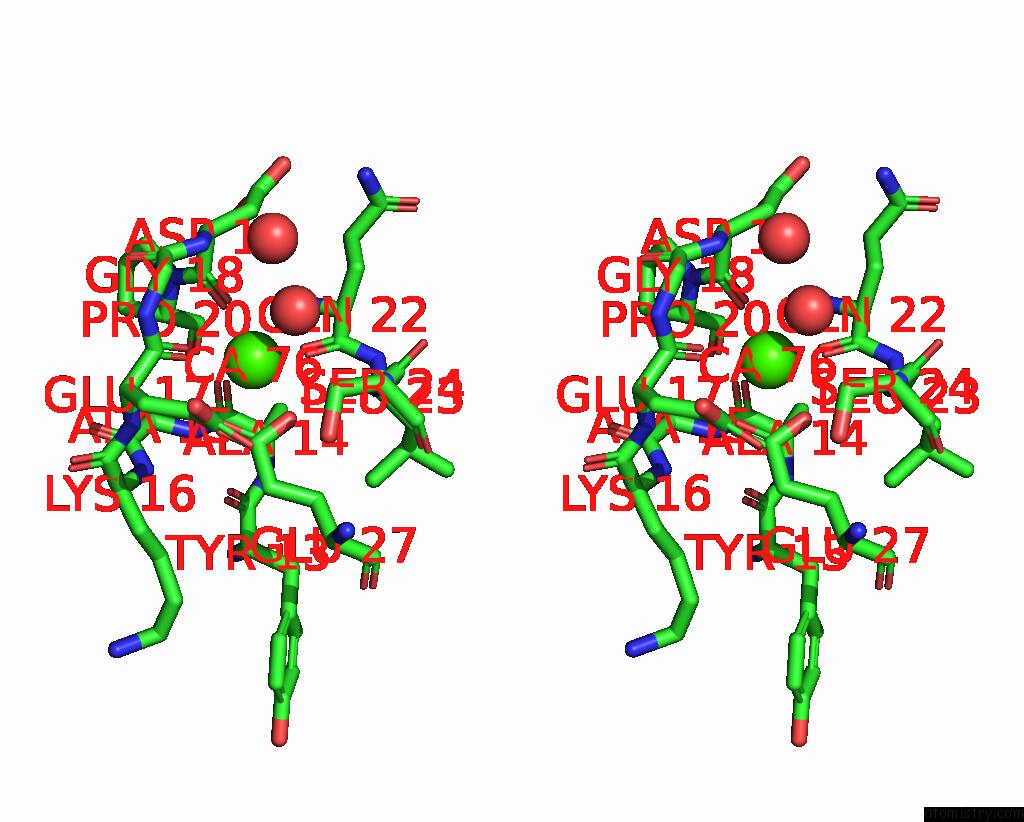
Stereo pair view
Mono view
Stereo pair view
A full contact list of Calcium with other atoms in the Ca binding
site number 1 of The Refined Structure of Vitamin D-Dependent Calcium- Binding Protein From Bovine Intestine. Molecular Details, Ion Binding, and Implications For the Structure of Other Calcium-Binding Proteins within 5.0Å range:
|
Calcium binding site 2 out of 2 in 3icb
Go back to
Calcium binding site 2 out
of 2 in the The Refined Structure of Vitamin D-Dependent Calcium- Binding Protein From Bovine Intestine. Molecular Details, Ion Binding, and Implications For the Structure of Other Calcium-Binding Proteins
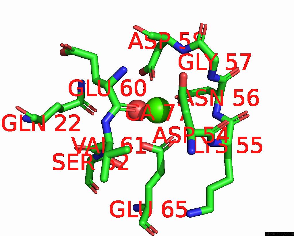
Mono view
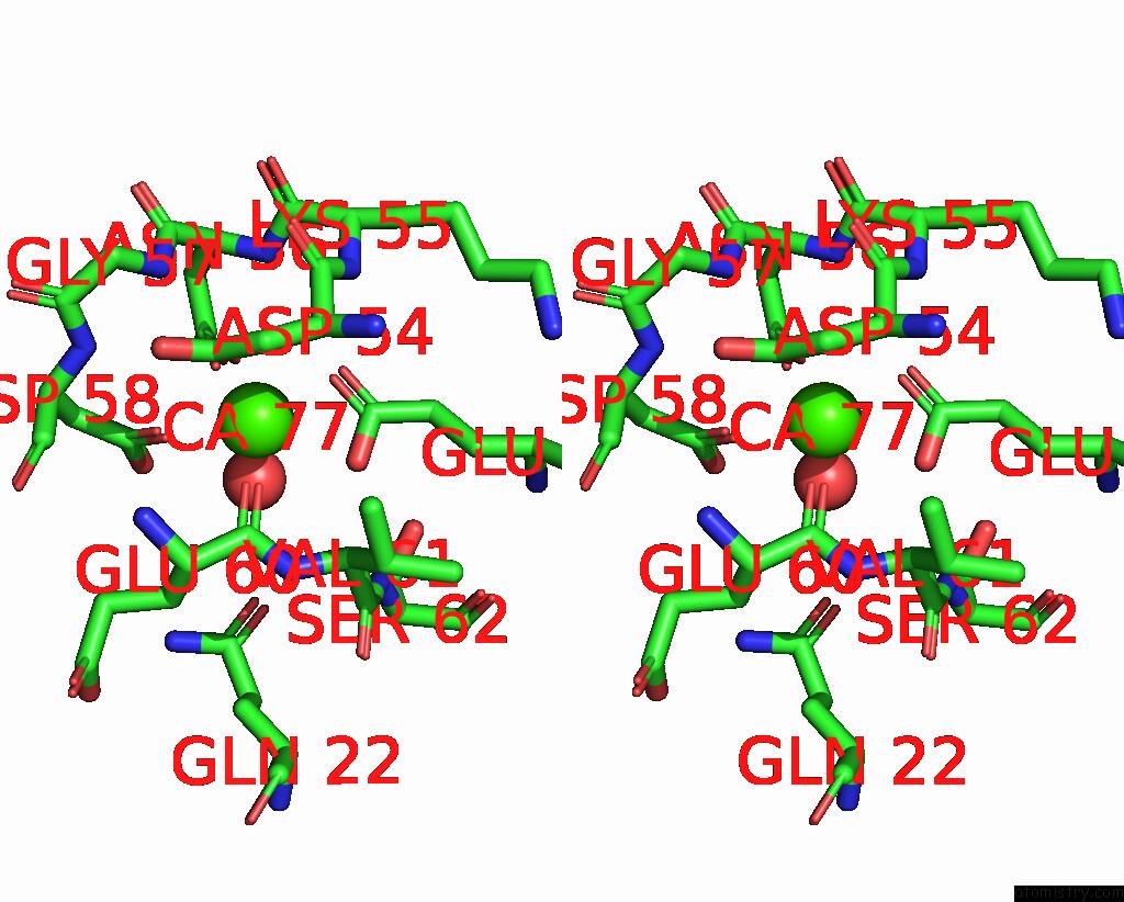
Stereo pair view
Mono view
Stereo pair view
A full contact list of Calcium with other atoms in the Ca binding
site number 2 of The Refined Structure of Vitamin D-Dependent Calcium- Binding Protein From Bovine Intestine. Molecular Details, Ion Binding, and Implications For the Structure of Other Calcium-Binding Proteins within 5.0Å range:
|
Reference:
D.M.Szebenyi,
K.Moffat.
The Refined Structure of Vitamin D-Dependent Calcium-Binding Protein From Bovine Intestine. Molecular Details, Ion Binding, and Implications For the Structure of Other Calcium-Binding Proteins. J.Biol.Chem. V. 261 8761 1986.
ISSN: ISSN 0021-9258
PubMed: 3722173
Page generated: Sat Jul 13 11:30:08 2024
ISSN: ISSN 0021-9258
PubMed: 3722173
Last articles
Zn in 9J0NZn in 9J0O
Zn in 9J0P
Zn in 9FJX
Zn in 9EKB
Zn in 9C0F
Zn in 9CAH
Zn in 9CH0
Zn in 9CH3
Zn in 9CH1