Calcium »
PDB 3ij7-3isv »
3iln »
Calcium in PDB 3iln: X-Ray Structure of the Laminarinase From Rhodothermus Marinus
Protein crystallography data
The structure of X-Ray Structure of the Laminarinase From Rhodothermus Marinus, PDB code: 3iln
was solved by
L.Bleicher,
A.Golubev,
A.L.Rojas,
A.S.Nascimento,
I.Polikarpov,
with X-Ray Crystallography technique. A brief refinement statistics is given in the table below:
| Resolution Low / High (Å) | 25.92 / 1.95 |
| Space group | P 1 21 1 |
| Cell size a, b, c (Å), α, β, γ (°) | 52.218, 108.288, 64.588, 90.00, 113.90, 90.00 |
| R / Rfree (%) | 15.7 / 19 |
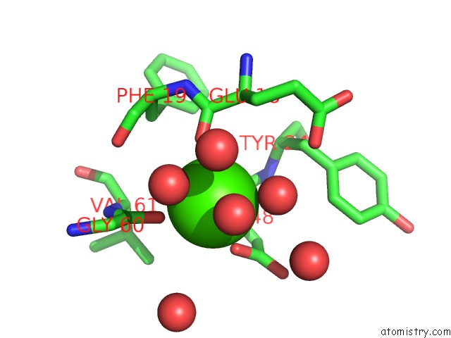
Calcium Binding Sites:
The binding sites of Calcium atom in the X-Ray Structure of the Laminarinase From Rhodothermus Marinus
(pdb code 3iln). This binding sites where shown within
5.0 Angstroms radius around Calcium atom.
In total 2 binding sites of Calcium where determined in the X-Ray Structure of the Laminarinase From Rhodothermus Marinus, PDB code: 3iln:
Jump to Calcium binding site number: 1; 2;
In total 2 binding sites of Calcium where determined in the X-Ray Structure of the Laminarinase From Rhodothermus Marinus, PDB code: 3iln:
Jump to Calcium binding site number: 1; 2;
Calcium binding site 1 out of 2 in 3iln
Go back to
Calcium binding site 1 out
of 2 in the X-Ray Structure of the Laminarinase From Rhodothermus Marinus

Mono view

Stereo pair view
Mono view
Stereo pair view
A full contact list of Calcium with other atoms in the Ca binding
site number 1 of X-Ray Structure of the Laminarinase From Rhodothermus Marinus within 5.0Å range:
|
Calcium binding site 2 out of 2 in 3iln
Go back to
Calcium binding site 2 out
of 2 in the X-Ray Structure of the Laminarinase From Rhodothermus Marinus
Mono view

Stereo pair view
Mono view
Stereo pair view
A full contact list of Calcium with other atoms in the Ca binding
site number 2 of X-Ray Structure of the Laminarinase From Rhodothermus Marinus within 5.0Å range:
|
Reference:
L.Bleicher,
E.T.Prates,
T.C.Gomes,
R.L.Silveira,
A.S.Nascimento,
A.L.Rojas,
A.Golubev,
L.Martinez,
M.S.Skaf,
I.Polikarpov.
Molecular Basis of the Thermostability and Thermophilicity of Laminarinases: X-Ray Structure of the Hyperthermostable Laminarinase From Rhodothermus Marinus and Molecular Dynamics Simulations. J.Phys.Chem.B V. 115 7940 2011.
ISSN: ISSN 1089-5647
PubMed: 21619042
DOI: 10.1021/JP200330Z
Page generated: Sat Jul 13 11:37:12 2024
ISSN: ISSN 1089-5647
PubMed: 21619042
DOI: 10.1021/JP200330Z
Last articles
Zn in 9JYWZn in 9IR4
Zn in 9IR3
Zn in 9GMX
Zn in 9GMW
Zn in 9JEJ
Zn in 9ERF
Zn in 9ERE
Zn in 9EGV
Zn in 9EGW