Calcium »
PDB 3ij7-3isv »
3ilr »
Calcium in PDB 3ilr: Structure of Heparinase I From Bacteroides Thetaiotaomicron in Complex with Tetrasaccharide Product
Enzymatic activity of Structure of Heparinase I From Bacteroides Thetaiotaomicron in Complex with Tetrasaccharide Product
All present enzymatic activity of Structure of Heparinase I From Bacteroides Thetaiotaomicron in Complex with Tetrasaccharide Product:
4.2.2.7;
4.2.2.7;
Protein crystallography data
The structure of Structure of Heparinase I From Bacteroides Thetaiotaomicron in Complex with Tetrasaccharide Product, PDB code: 3ilr
was solved by
M.L.Garron,
M.Cygler,
D.Shaya,
with X-Ray Crystallography technique. A brief refinement statistics is given in the table below:
| Resolution Low / High (Å) | 19.81 / 1.50 |
| Space group | P 21 21 2 |
| Cell size a, b, c (Å), α, β, γ (°) | 75.736, 109.604, 43.858, 90.00, 90.00, 90.00 |
| R / Rfree (%) | 17.6 / 20.9 |
Calcium Binding Sites:
The binding sites of Calcium atom in the Structure of Heparinase I From Bacteroides Thetaiotaomicron in Complex with Tetrasaccharide Product
(pdb code 3ilr). This binding sites where shown within
5.0 Angstroms radius around Calcium atom.
In total only one binding site of Calcium was determined in the Structure of Heparinase I From Bacteroides Thetaiotaomicron in Complex with Tetrasaccharide Product, PDB code: 3ilr:
In total only one binding site of Calcium was determined in the Structure of Heparinase I From Bacteroides Thetaiotaomicron in Complex with Tetrasaccharide Product, PDB code: 3ilr:
Calcium binding site 1 out of 1 in 3ilr
Go back to
Calcium binding site 1 out
of 1 in the Structure of Heparinase I From Bacteroides Thetaiotaomicron in Complex with Tetrasaccharide Product

Mono view
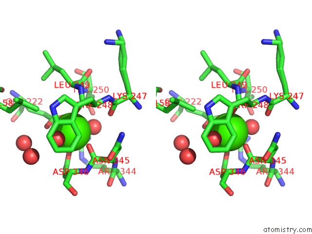
Stereo pair view
Mono view
Stereo pair view
A full contact list of Calcium with other atoms in the Ca binding
site number 1 of Structure of Heparinase I From Bacteroides Thetaiotaomicron in Complex with Tetrasaccharide Product within 5.0Å range:
|
Reference:
Y.H.Han,
M.L.Garron,
H.Y.Kim,
W.S.Kim,
Z.Zhang,
K.S.Ryu,
D.Shaya,
Z.Xiao,
C.Cheong,
Y.S.Kim,
R.J.Linhardt,
Y.H.Jeon,
M.Cygler.
Structural Snapshots of Heparin Depolymerization By Heparin Lyase I. J.Biol.Chem. V. 284 34019 2009.
ISSN: ISSN 0021-9258
PubMed: 19801541
DOI: 10.1074/JBC.M109.025338
Page generated: Sat Jul 13 11:37:42 2024
ISSN: ISSN 0021-9258
PubMed: 19801541
DOI: 10.1074/JBC.M109.025338
Last articles
Zn in 9MJ5Zn in 9HNW
Zn in 9G0L
Zn in 9FNE
Zn in 9DZN
Zn in 9E0I
Zn in 9D32
Zn in 9DAK
Zn in 8ZXC
Zn in 8ZUF