Calcium »
PDB 3ij7-3isv »
3iqo »
Calcium in PDB 3iqo: 1.5 Angstrom X-Ray Structure of Bovine Ca(2+)-S100B
Protein crystallography data
The structure of 1.5 Angstrom X-Ray Structure of Bovine Ca(2+)-S100B, PDB code: 3iqo
was solved by
T.H.Charpentier,
D.J.Weber,
E.A.Toth,
with X-Ray Crystallography technique. A brief refinement statistics is given in the table below:
| Resolution Low / High (Å) | 32.63 / 1.50 |
| Space group | C 1 2 1 |
| Cell size a, b, c (Å), α, β, γ (°) | 89.637, 35.044, 58.113, 90.00, 92.62, 90.00 |
| R / Rfree (%) | 20 / 24.2 |
Calcium Binding Sites:
The binding sites of Calcium atom in the 1.5 Angstrom X-Ray Structure of Bovine Ca(2+)-S100B
(pdb code 3iqo). This binding sites where shown within
5.0 Angstroms radius around Calcium atom.
In total 4 binding sites of Calcium where determined in the 1.5 Angstrom X-Ray Structure of Bovine Ca(2+)-S100B, PDB code: 3iqo:
Jump to Calcium binding site number: 1; 2; 3; 4;
In total 4 binding sites of Calcium where determined in the 1.5 Angstrom X-Ray Structure of Bovine Ca(2+)-S100B, PDB code: 3iqo:
Jump to Calcium binding site number: 1; 2; 3; 4;
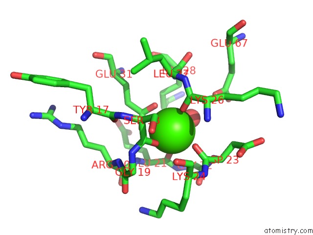
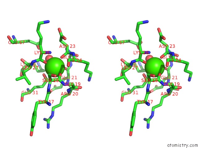
Calcium binding site 1 out of 4 in 3iqo
Go back to
Calcium binding site 1 out
of 4 in the 1.5 Angstrom X-Ray Structure of Bovine Ca(2+)-S100B
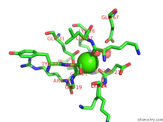
Mono view
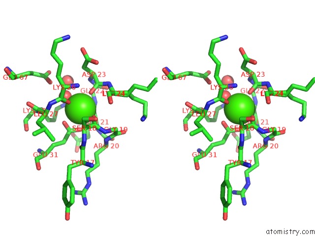
Stereo pair view
Mono view
Stereo pair view
A full contact list of Calcium with other atoms in the Ca binding
site number 1 of 1.5 Angstrom X-Ray Structure of Bovine Ca(2+)-S100B within 5.0Å range:
|
Calcium binding site 2 out of 4 in 3iqo
Go back to
Calcium binding site 2 out
of 4 in the 1.5 Angstrom X-Ray Structure of Bovine Ca(2+)-S100B
Mono view
Stereo pair view
Mono view
Stereo pair view
A full contact list of Calcium with other atoms in the Ca binding
site number 2 of 1.5 Angstrom X-Ray Structure of Bovine Ca(2+)-S100B within 5.0Å range:
|
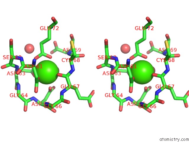
Calcium binding site 3 out of 4 in 3iqo
Go back to
Calcium binding site 3 out
of 4 in the 1.5 Angstrom X-Ray Structure of Bovine Ca(2+)-S100B
Mono view
Stereo pair view
Mono view
Stereo pair view
A full contact list of Calcium with other atoms in the Ca binding
site number 3 of 1.5 Angstrom X-Ray Structure of Bovine Ca(2+)-S100B within 5.0Å range:
|
Calcium binding site 4 out of 4 in 3iqo
Go back to
Calcium binding site 4 out
of 4 in the 1.5 Angstrom X-Ray Structure of Bovine Ca(2+)-S100B
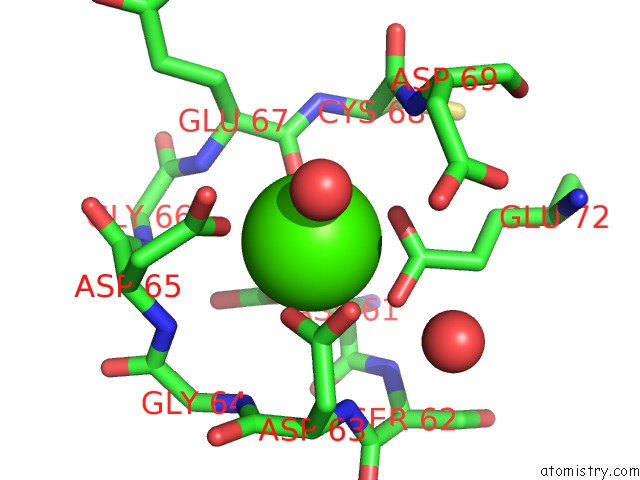
Mono view

Stereo pair view
Mono view
Stereo pair view
A full contact list of Calcium with other atoms in the Ca binding
site number 4 of 1.5 Angstrom X-Ray Structure of Bovine Ca(2+)-S100B within 5.0Å range:
|
Reference:
T.H.Charpentier,
L.E.Thompson,
M.A.Liriano,
K.M.Varney,
P.T.Wilder,
E.Pozharski,
E.A.Toth,
D.J.Weber.
The Effects of Capz Peptide (Trtk-12) Binding to S100B-Ca(2+) As Examined By uc(Nmr) and X-Ray Crystallography J.Mol.Biol. V. 396 1227 2010.
ISSN: ISSN 0022-2836
PubMed: 20053360
DOI: 10.1016/J.JMB.2009.12.057
Page generated: Tue Jul 8 13:28:08 2025
ISSN: ISSN 0022-2836
PubMed: 20053360
DOI: 10.1016/J.JMB.2009.12.057
Last articles
Cl in 8DUZCl in 8DTQ
Cl in 8DT6
Cl in 8DSZ
Cl in 8DTE
Cl in 8DTJ
Cl in 8DSR
Cl in 8DSY
Cl in 8DSM
Cl in 8DSI