Calcium in PDB 3iqq: X-Ray Structure of Bovine TRTK12-Ca(2+)-S100B
Protein crystallography data
The structure of X-Ray Structure of Bovine TRTK12-Ca(2+)-S100B, PDB code: 3iqq
was solved by
T.H.Charpentier,
D.J.Weber,
E.A.Toth,
with X-Ray Crystallography technique. A brief refinement statistics is given in the table below:
Resolution Low / High (Å)
|
35.69 /
2.01
|
Space group
|
C 2 2 21
|
Cell size a, b, c (Å), α, β, γ (°)
|
32.951,
89.772,
58.860,
90.00,
90.00,
90.00
|
R / Rfree (%)
|
22.1 /
26.6
|
Calcium Binding Sites:
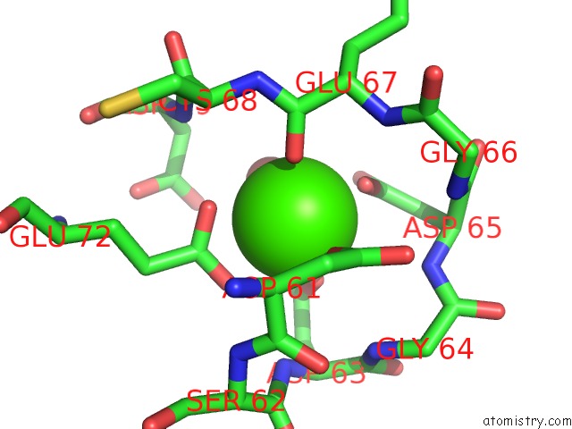
The binding sites of Calcium atom in the X-Ray Structure of Bovine TRTK12-Ca(2+)-S100B
(pdb code 3iqq). This binding sites where shown within
5.0 Angstroms radius around Calcium atom.
In total 2 binding sites of Calcium where determined in the
X-Ray Structure of Bovine TRTK12-Ca(2+)-S100B, PDB code: 3iqq:
Jump to Calcium binding site number:
1;
2;
Calcium binding site 1 out
of 2 in 3iqq
Go back to
Calcium Binding Sites List in 3iqq
Calcium binding site 1 out
of 2 in the X-Ray Structure of Bovine TRTK12-Ca(2+)-S100B
 Mono view
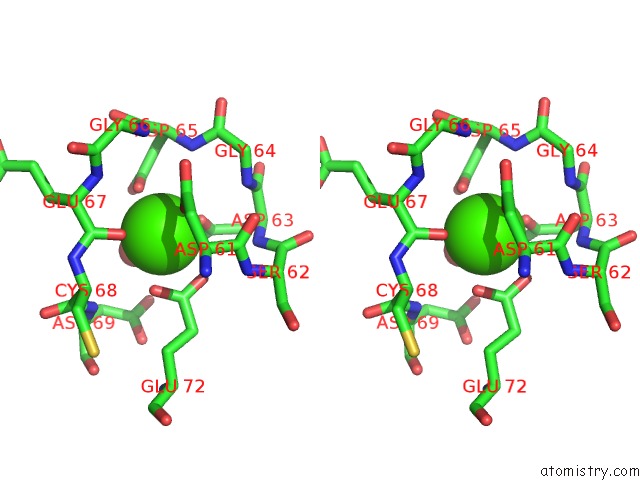
 Stereo pair view
|
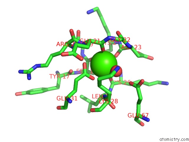
A full contact list of Calcium with other atoms in the Ca binding
site number 1 of X-Ray Structure of Bovine TRTK12-Ca(2+)-S100B within 5.0Å range:
probe
|
atom
|
residue
|
distance (Å)
|
B
|
Occ
|
A:Ca92
b:32.5
occ:1.00
|
OD1
|
A:ASP61
|
2.2
|
35.3
|
1.0
|
O
|
A:GLU67
|
2.3
|
33.8
|
1.0
|
OD1
|
A:ASP63
|
2.4
|
30.7
|
1.0
|
OD1
|
A:ASP65
|
2.4
|
32.9
|
1.0
|
O
|
A:HOH98
|
2.4
|
37.2
|
1.0
|
OE1
|
A:GLU72
|
2.5
|
32.3
|
1.0
|
OE2
|
A:GLU72
|
2.7
|
30.9
|
1.0
|
CD
|
A:GLU72
|
2.9
|
33.0
|
1.0
|
CG
|
A:ASP65
|
3.3
|
35.9
|
1.0
|
CG
|
A:ASP63
|
3.4
|
33.3
|
1.0
|
CG
|
A:ASP61
|
3.4
|
35.1
|
1.0
|
C
|
A:GLU67
|
3.5
|
34.5
|
1.0
|
OD2
|
A:ASP65
|
3.7
|
39.8
|
1.0
|
OD2
|
A:ASP63
|
3.8
|
34.5
|
1.0
|
N
|
A:ASP65
|
4.1
|
33.7
|
1.0
|
CA
|
A:ASP61
|
4.1
|
33.6
|
1.0
|
N
|
A:GLU67
|
4.2
|
34.1
|
1.0
|
CB
|
A:ASP61
|
4.2
|
33.4
|
1.0
|
OD2
|
A:ASP61
|
4.2
|
34.5
|
1.0
|
N
|
A:ASP63
|
4.3
|
33.0
|
1.0
|
N
|
A:GLY64
|
4.4
|
33.1
|
1.0
|
CB
|
A:ASP65
|
4.4
|
34.8
|
1.0
|
N
|
A:CYS68
|
4.4
|
33.8
|
1.0
|
CA
|
A:GLU67
|
4.4
|
34.6
|
1.0
|
C
|
A:ASP61
|
4.4
|
33.5
|
1.0
|
N
|
A:ASP69
|
4.4
|
34.4
|
1.0
|
CG
|
A:GLU72
|
4.4
|
31.4
|
1.0
|
CA
|
A:CYS68
|
4.5
|
34.6
|
1.0
|
N
|
A:SER62
|
4.6
|
33.3
|
1.0
|
OD2
|
A:ASP69
|
4.6
|
33.7
|
1.0
|
CA
|
A:ASP65
|
4.6
|
34.4
|
1.0
|
CB
|
A:ASP63
|
4.6
|
33.5
|
1.0
|
N
|
A:GLY66
|
4.8
|
34.2
|
1.0
|
CA
|
A:ASP63
|
4.8
|
33.4
|
1.0
|
C
|
A:ASP63
|
4.8
|
33.1
|
1.0
|
CG
|
A:ASP69
|
4.8
|
34.5
|
1.0
|
C
|
A:ASP65
|
4.9
|
34.6
|
1.0
|
C
|
A:CYS68
|
5.0
|
34.2
|
1.0
|
O
|
A:ASP61
|
5.0
|
32.8
|
1.0
|
|
Calcium binding site 2 out
of 2 in 3iqq
Go back to
Calcium Binding Sites List in 3iqq
Calcium binding site 2 out
of 2 in the X-Ray Structure of Bovine TRTK12-Ca(2+)-S100B
 Mono view
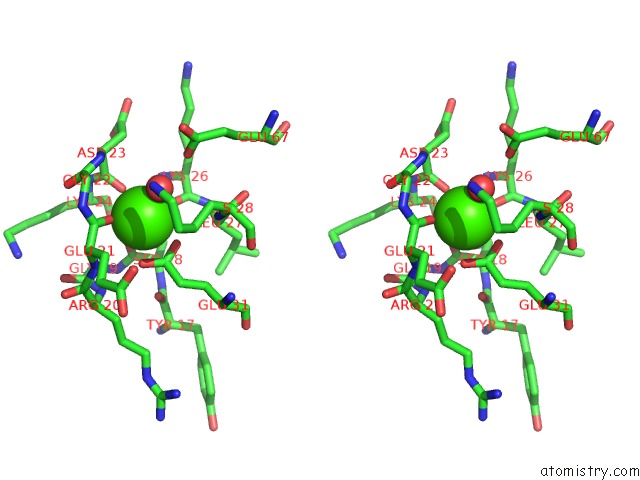
 Stereo pair view
|
A full contact list of Calcium with other atoms in the Ca binding
site number 2 of X-Ray Structure of Bovine TRTK12-Ca(2+)-S100B within 5.0Å range:
probe
|
atom
|
residue
|
distance (Å)
|
B
|
Occ
|
A:Ca93
b:31.5
occ:1.00
|
O
|
A:HOH104
|
2.3
|
32.6
|
1.0
|
O
|
A:SER18
|
2.3
|
32.2
|
1.0
|
O
|
A:GLU21
|
2.3
|
33.8
|
1.0
|
OE1
|
A:GLU31
|
2.4
|
34.4
|
1.0
|
O
|
A:ASP23
|
2.4
|
32.8
|
1.0
|
O
|
A:LYS26
|
2.4
|
34.6
|
1.0
|
OE2
|
A:GLU31
|
2.7
|
30.6
|
1.0
|
CD
|
A:GLU31
|
2.9
|
32.5
|
1.0
|
C
|
A:SER18
|
3.4
|
32.5
|
1.0
|
C
|
A:ASP23
|
3.5
|
32.7
|
1.0
|
C
|
A:GLU21
|
3.6
|
34.0
|
1.0
|
C
|
A:LYS26
|
3.7
|
34.3
|
1.0
|
CA
|
A:SER18
|
4.0
|
33.3
|
1.0
|
N
|
A:GLU21
|
4.1
|
33.4
|
1.0
|
N
|
A:ASP23
|
4.1
|
32.6
|
1.0
|
C
|
A:GLY22
|
4.2
|
33.0
|
1.0
|
N
|
A:LYS28
|
4.3
|
32.9
|
1.0
|
OE1
|
A:GLU67
|
4.4
|
37.0
|
1.0
|
CA
|
A:ASP23
|
4.4
|
32.5
|
1.0
|
CG
|
A:GLU31
|
4.4
|
31.4
|
1.0
|
CA
|
A:GLU21
|
4.4
|
34.9
|
1.0
|
CA
|
A:LEU27
|
4.4
|
33.3
|
1.0
|
N
|
A:LYS24
|
4.4
|
33.1
|
1.0
|
N
|
A:LEU27
|
4.5
|
34.0
|
1.0
|
CA
|
A:LYS24
|
4.5
|
33.9
|
1.0
|
N
|
A:GLY22
|
4.5
|
33.5
|
1.0
|
N
|
A:GLY19
|
4.5
|
31.9
|
1.0
|
O
|
A:GLY22
|
4.6
|
32.8
|
1.0
|
N
|
A:LYS26
|
4.6
|
34.7
|
1.0
|
CA
|
A:GLY22
|
4.6
|
33.2
|
1.0
|
CG
|
A:LYS28
|
4.6
|
34.5
|
1.0
|
CB
|
A:SER18
|
4.6
|
32.9
|
1.0
|
O
|
A:TYR17
|
4.7
|
33.5
|
1.0
|
CA
|
A:LYS26
|
4.7
|
34.1
|
1.0
|
N
|
A:ARG20
|
4.7
|
32.8
|
1.0
|
C
|
A:LEU27
|
4.7
|
33.5
|
1.0
|
CB
|
A:ASP23
|
4.8
|
32.0
|
1.0
|
C
|
A:LYS24
|
4.9
|
34.0
|
1.0
|
CA
|
A:GLY19
|
4.9
|
32.3
|
1.0
|
C
|
A:GLY19
|
5.0
|
32.5
|
1.0
|
CB
|
A:GLU21
|
5.0
|
35.2
|
1.0
|
|
Reference:
T.H.Charpentier,
L.E.Thompson,
M.A.Liriano,
K.M.Varney,
P.T.Wilder,
E.Pozharski,
E.A.Toth,
D.J.Weber.
The Effects of Capz Peptide (Trtk-12) Binding to S100B-Ca(2+) As Examined By uc(Nmr) and X-Ray Crystallography J.Mol.Biol. V. 396 1227 2010.
ISSN: ISSN 0022-2836
PubMed: 20053360
DOI: 10.1016/J.JMB.2009.12.057
Page generated: Sat Jul 13 11:45:46 2024
|