Calcium »
PDB 3ij7-3isv »
3iqt »
Calcium in PDB 3iqt: Structure of the Hpt Domain of Sensor Protein Bara From Escherichia Coli CFT073.
Enzymatic activity of Structure of the Hpt Domain of Sensor Protein Bara From Escherichia Coli CFT073.
All present enzymatic activity of Structure of the Hpt Domain of Sensor Protein Bara From Escherichia Coli CFT073.:
2.7.13.3;
2.7.13.3;
Protein crystallography data
The structure of Structure of the Hpt Domain of Sensor Protein Bara From Escherichia Coli CFT073., PDB code: 3iqt
was solved by
M.E.Cuff,
E.Rakowski,
Y.Kim,
L.Freeman,
A.Joachimiak,
Midwest Center Forstructural Genomics (Mcsg),
with X-Ray Crystallography technique. A brief refinement statistics is given in the table below:
| Resolution Low / High (Å) | 35.30 / 1.40 |
| Space group | P 1 21 1 |
| Cell size a, b, c (Å), α, β, γ (°) | 24.316, 60.359, 37.156, 90.00, 108.21, 90.00 |
| R / Rfree (%) | 14.1 / 18.1 |
Calcium Binding Sites:
The binding sites of Calcium atom in the Structure of the Hpt Domain of Sensor Protein Bara From Escherichia Coli CFT073.
(pdb code 3iqt). This binding sites where shown within
5.0 Angstroms radius around Calcium atom.
In total only one binding site of Calcium was determined in the Structure of the Hpt Domain of Sensor Protein Bara From Escherichia Coli CFT073., PDB code: 3iqt:
In total only one binding site of Calcium was determined in the Structure of the Hpt Domain of Sensor Protein Bara From Escherichia Coli CFT073., PDB code: 3iqt:
Calcium binding site 1 out of 1 in 3iqt
Go back to
Calcium binding site 1 out
of 1 in the Structure of the Hpt Domain of Sensor Protein Bara From Escherichia Coli CFT073.
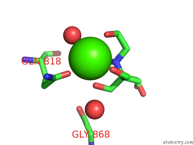
Mono view
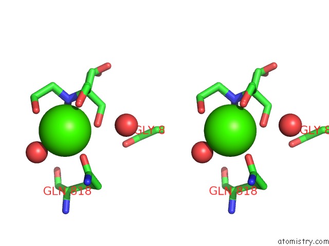
Stereo pair view
Mono view
Stereo pair view
A full contact list of Calcium with other atoms in the Ca binding
site number 1 of Structure of the Hpt Domain of Sensor Protein Bara From Escherichia Coli CFT073. within 5.0Å range:
|
Reference:
M.E.Cuff,
E.Rakowski,
Y.Kim,
L.Freeman,
A.Joachimiak.
Structure of the Hpt Domain of Sensor Protein Bara From Escherichia Coli CFT073. To Be Published.
Page generated: Tue Jul 8 13:28:58 2025
Last articles
Cl in 5K0TCl in 5K0B
Cl in 5JZS
Cl in 5K0E
Cl in 5JY3
Cl in 5JZN
Cl in 5JZB
Cl in 5JZL
Cl in 5JZ9
Cl in 5JZK