Calcium »
PDB 3iti-3k3w »
3iti »
Calcium in PDB 3iti: Structure of Bovine Trypsin with the Mad Triangle B3C
Enzymatic activity of Structure of Bovine Trypsin with the Mad Triangle B3C
All present enzymatic activity of Structure of Bovine Trypsin with the Mad Triangle B3C:
3.4.21.4;
3.4.21.4;
Protein crystallography data
The structure of Structure of Bovine Trypsin with the Mad Triangle B3C, PDB code: 3iti
was solved by
T.Beck,
C.E.Da Cunha,
G.M.Sheldrick,
with X-Ray Crystallography technique. A brief refinement statistics is given in the table below:
| Resolution Low / High (Å) | 41.84 / 1.55 |
| Space group | P 21 21 21 |
| Cell size a, b, c (Å), α, β, γ (°) | 53.658, 56.883, 66.808, 90.00, 90.00, 90.00 |
| R / Rfree (%) | 16.8 / 21.3 |
Other elements in 3iti:
The structure of Structure of Bovine Trypsin with the Mad Triangle B3C also contains other interesting chemical elements:
| Bromine | (Br) | 3 atoms |
| Chlorine | (Cl) | 1 atom |
Calcium Binding Sites:
The binding sites of Calcium atom in the Structure of Bovine Trypsin with the Mad Triangle B3C
(pdb code 3iti). This binding sites where shown within
5.0 Angstroms radius around Calcium atom.
In total only one binding site of Calcium was determined in the Structure of Bovine Trypsin with the Mad Triangle B3C, PDB code: 3iti:
In total only one binding site of Calcium was determined in the Structure of Bovine Trypsin with the Mad Triangle B3C, PDB code: 3iti:
Calcium binding site 1 out of 1 in 3iti
Go back to
Calcium binding site 1 out
of 1 in the Structure of Bovine Trypsin with the Mad Triangle B3C
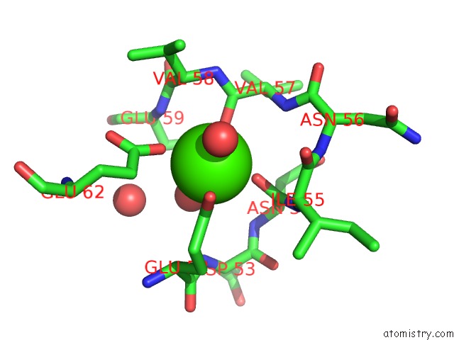
Mono view
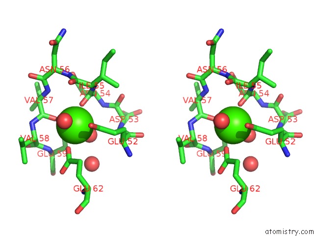
Stereo pair view
Mono view
Stereo pair view
A full contact list of Calcium with other atoms in the Ca binding
site number 1 of Structure of Bovine Trypsin with the Mad Triangle B3C within 5.0Å range:
|
Reference:
T.Beck,
C.E.Da Cunha,
G.M.Sheldrick.
How to Get the Magic Triangle and the Mad Triangle Into Your Protein Crystal. Acta Crystallogr.,Sect.F V. 65 1068 2009.
ISSN: ESSN 1744-3091
PubMed: 19851024
DOI: 10.1107/S1744309109036884
Page generated: Sat Jul 13 11:49:11 2024
ISSN: ESSN 1744-3091
PubMed: 19851024
DOI: 10.1107/S1744309109036884
Last articles
Zn in 9J0NZn in 9J0O
Zn in 9J0P
Zn in 9FJX
Zn in 9EKB
Zn in 9C0F
Zn in 9CAH
Zn in 9CH0
Zn in 9CH3
Zn in 9CH1