Calcium »
PDB 3iti-3k3w »
3j7t »
Calcium in PDB 3j7t: Calcium Atpase Structure with Two Bound Calcium Ions Determined By Electron Crystallography of Thin 3D Crystals
Enzymatic activity of Calcium Atpase Structure with Two Bound Calcium Ions Determined By Electron Crystallography of Thin 3D Crystals
All present enzymatic activity of Calcium Atpase Structure with Two Bound Calcium Ions Determined By Electron Crystallography of Thin 3D Crystals:
3.6.3.8;
3.6.3.8;
Other elements in 3j7t:
The structure of Calcium Atpase Structure with Two Bound Calcium Ions Determined By Electron Crystallography of Thin 3D Crystals also contains other interesting chemical elements:
| Sodium | (Na) | 1 atom |
Calcium Binding Sites:
The binding sites of Calcium atom in the Calcium Atpase Structure with Two Bound Calcium Ions Determined By Electron Crystallography of Thin 3D Crystals
(pdb code 3j7t). This binding sites where shown within
5.0 Angstroms radius around Calcium atom.
In total 2 binding sites of Calcium where determined in the Calcium Atpase Structure with Two Bound Calcium Ions Determined By Electron Crystallography of Thin 3D Crystals, PDB code: 3j7t:
Jump to Calcium binding site number: 1; 2;
In total 2 binding sites of Calcium where determined in the Calcium Atpase Structure with Two Bound Calcium Ions Determined By Electron Crystallography of Thin 3D Crystals, PDB code: 3j7t:
Jump to Calcium binding site number: 1; 2;
Calcium binding site 1 out of 2 in 3j7t
Go back to
Calcium binding site 1 out
of 2 in the Calcium Atpase Structure with Two Bound Calcium Ions Determined By Electron Crystallography of Thin 3D Crystals
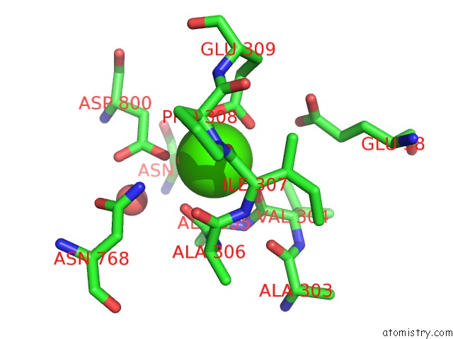
Mono view
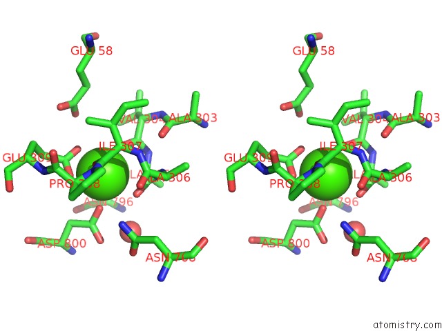
Stereo pair view
Mono view
Stereo pair view
A full contact list of Calcium with other atoms in the Ca binding
site number 1 of Calcium Atpase Structure with Two Bound Calcium Ions Determined By Electron Crystallography of Thin 3D Crystals within 5.0Å range:
|
Calcium binding site 2 out of 2 in 3j7t
Go back to
Calcium binding site 2 out
of 2 in the Calcium Atpase Structure with Two Bound Calcium Ions Determined By Electron Crystallography of Thin 3D Crystals
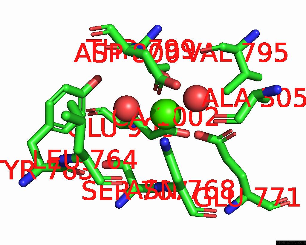
Mono view
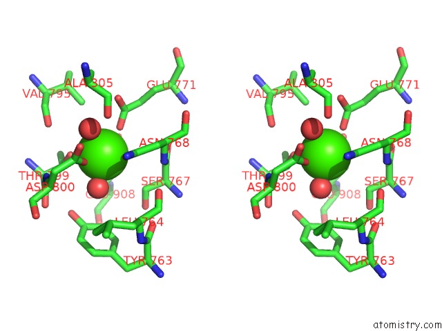
Stereo pair view
Mono view
Stereo pair view
A full contact list of Calcium with other atoms in the Ca binding
site number 2 of Calcium Atpase Structure with Two Bound Calcium Ions Determined By Electron Crystallography of Thin 3D Crystals within 5.0Å range:
|
Reference:
K.Yonekura,
K.Kato,
M.Ogasawara,
M.Tomita,
C.Toyoshima.
Electron Crystallography of Ultra-Thin 3D Protein Crystals: Atomic Model with Charges Proc.Natl.Acad.Sci.Usa 2015.
ISSN: ESSN 1091-6490
Page generated: Sat Jul 13 11:49:11 2024
ISSN: ESSN 1091-6490
Last articles
Zn in 9J0NZn in 9J0O
Zn in 9J0P
Zn in 9FJX
Zn in 9EKB
Zn in 9C0F
Zn in 9CAH
Zn in 9CH0
Zn in 9CH3
Zn in 9CH1