Calcium in PDB 3k21: Crystal Structure of Carboxy-Terminus of PFC0420W.
Enzymatic activity of Crystal Structure of Carboxy-Terminus of PFC0420W.
All present enzymatic activity of Crystal Structure of Carboxy-Terminus of PFC0420W.:
2.7.11.1;
Protein crystallography data
The structure of Crystal Structure of Carboxy-Terminus of PFC0420W., PDB code: 3k21
was solved by
A.K.Wernimont,
A.Hutchinson,
J.D.Artz,
F.Mackenzie,
D.Cossar,
I.Kozieradzki,
C.H.Arrowsmith,
A.M.Edwards,
C.Bountra,
J.Weigelt,
A.Bochkarev,
R.Hui,
M.Amani,
Structural Genomics Consortium (Sgc),
with X-Ray Crystallography technique. A brief refinement statistics is given in the table below:
Resolution Low / High (Å)
|
16.73 /
1.15
|
Space group
|
P 31 2 1
|
Cell size a, b, c (Å), α, β, γ (°)
|
50.469,
50.469,
108.656,
90.00,
90.00,
120.00
|
R / Rfree (%)
|
15 /
18.6
|
Calcium Binding Sites:
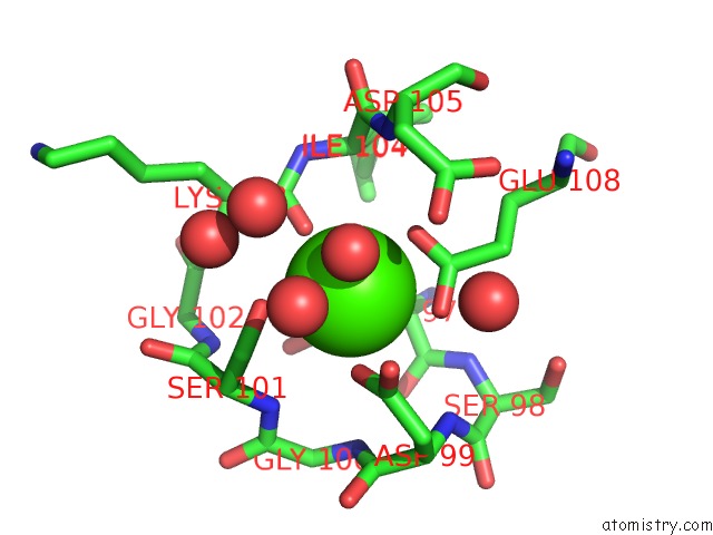
The binding sites of Calcium atom in the Crystal Structure of Carboxy-Terminus of PFC0420W.
(pdb code 3k21). This binding sites where shown within
5.0 Angstroms radius around Calcium atom.
In total 3 binding sites of Calcium where determined in the
Crystal Structure of Carboxy-Terminus of PFC0420W., PDB code: 3k21:
Jump to Calcium binding site number:
1;
2;
3;
Calcium binding site 1 out
of 3 in 3k21
Go back to
Calcium Binding Sites List in 3k21
Calcium binding site 1 out
of 3 in the Crystal Structure of Carboxy-Terminus of PFC0420W.
Mono view
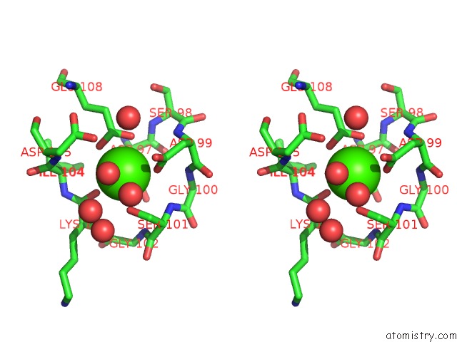
Stereo pair view
|
A full contact list of Calcium with other atoms in the Ca binding
site number 1 of Crystal Structure of Carboxy-Terminus of PFC0420W. within 5.0Å range:
probe
|
atom
|
residue
|
distance (Å)
|
B
|
Occ
|
A:Ca192
b:11.1
occ:1.00
|
OD1
|
A:ASP97
|
2.2
|
11.4
|
1.0
|
O
|
A:HOH226
|
2.3
|
18.0
|
1.0
|
OD1
|
A:ASP99
|
2.4
|
15.1
|
1.0
|
O
|
A:LYS103
|
2.4
|
12.0
|
1.0
|
OE1
|
A:GLU108
|
2.4
|
12.2
|
1.0
|
OE2
|
A:GLU108
|
2.5
|
12.7
|
1.0
|
OG
|
A:SER101
|
2.5
|
14.3
|
1.0
|
CD
|
A:GLU108
|
2.8
|
10.8
|
1.0
|
CG
|
A:ASP99
|
3.3
|
15.9
|
1.0
|
CG
|
A:ASP97
|
3.4
|
10.8
|
1.0
|
CB
|
A:SER101
|
3.5
|
16.0
|
1.0
|
OD2
|
A:ASP99
|
3.6
|
19.8
|
1.0
|
C
|
A:LYS103
|
3.6
|
10.3
|
1.0
|
N
|
A:SER101
|
3.9
|
14.7
|
1.0
|
CA
|
A:SER101
|
4.2
|
14.9
|
1.0
|
OD2
|
A:ASP97
|
4.2
|
13.3
|
1.0
|
CA
|
A:ASP97
|
4.2
|
8.5
|
1.0
|
CB
|
A:ASP97
|
4.3
|
9.2
|
1.0
|
CG
|
A:GLU108
|
4.3
|
9.9
|
1.0
|
N
|
A:LYS103
|
4.3
|
11.9
|
1.0
|
O
|
A:HOH365
|
4.4
|
61.8
|
1.0
|
O
|
A:HOH355
|
4.4
|
21.0
|
1.0
|
CA
|
A:ILE104
|
4.4
|
8.5
|
0.5
|
N
|
A:ASP105
|
4.4
|
9.3
|
1.0
|
CA
|
A:ILE104
|
4.5
|
9.0
|
0.5
|
N
|
A:ILE104
|
4.5
|
9.5
|
1.0
|
C
|
A:ASP97
|
4.5
|
10.5
|
1.0
|
N
|
A:ASP99
|
4.5
|
12.6
|
1.0
|
CB
|
A:ASP99
|
4.5
|
15.1
|
1.0
|
OD2
|
A:ASP105
|
4.6
|
26.1
|
1.0
|
O
|
A:HOH354
|
4.6
|
27.0
|
1.0
|
N
|
A:GLY102
|
4.6
|
14.6
|
1.0
|
CA
|
A:LYS103
|
4.6
|
11.7
|
1.0
|
N
|
A:GLY100
|
4.6
|
13.4
|
1.0
|
C
|
A:SER101
|
4.8
|
15.2
|
1.0
|
O
|
A:HOH378
|
4.8
|
19.7
|
0.6
|
CA
|
A:ASP99
|
4.9
|
13.6
|
1.0
|
C
|
A:ASP99
|
4.9
|
12.6
|
1.0
|
N
|
A:SER98
|
4.9
|
10.5
|
1.0
|
O
|
A:ASP97
|
4.9
|
12.3
|
1.0
|
CG
|
A:ASP105
|
4.9
|
17.3
|
1.0
|
C
|
A:ILE104
|
4.9
|
9.3
|
1.0
|
C
|
A:GLY100
|
5.0
|
16.9
|
1.0
|
|
Calcium binding site 2 out
of 3 in 3k21
Go back to
Calcium Binding Sites List in 3k21
Calcium binding site 2 out
of 3 in the Crystal Structure of Carboxy-Terminus of PFC0420W.
Mono view
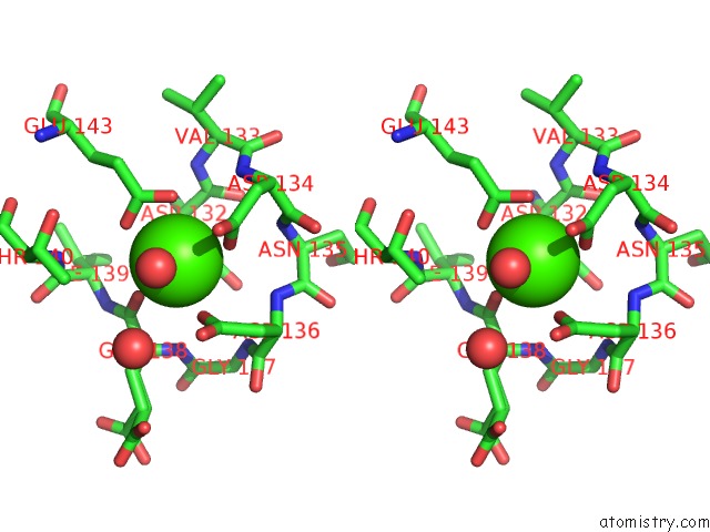
Stereo pair view
|
A full contact list of Calcium with other atoms in the Ca binding
site number 2 of Crystal Structure of Carboxy-Terminus of PFC0420W. within 5.0Å range:
probe
|
atom
|
residue
|
distance (Å)
|
B
|
Occ
|
A:Ca193
b:7.3
occ:1.00
|
OD1
|
A:ASP132
|
2.3
|
8.3
|
1.0
|
O
|
A:GLU138
|
2.3
|
10.2
|
1.0
|
OD1
|
A:ASP134
|
2.4
|
8.8
|
1.0
|
OD1
|
A:ASP136
|
2.4
|
9.0
|
1.0
|
OE1
|
A:GLU143
|
2.4
|
9.3
|
1.0
|
O
|
A:HOH236
|
2.4
|
13.3
|
1.0
|
OE2
|
A:GLU143
|
2.5
|
9.2
|
1.0
|
CD
|
A:GLU143
|
2.8
|
7.7
|
1.0
|
CG
|
A:ASP134
|
3.3
|
9.2
|
1.0
|
CG
|
A:ASP136
|
3.3
|
10.0
|
1.0
|
CG
|
A:ASP132
|
3.5
|
6.8
|
1.0
|
C
|
A:GLU138
|
3.5
|
7.4
|
1.0
|
OD2
|
A:ASP134
|
3.8
|
10.7
|
1.0
|
OD2
|
A:ASP136
|
3.8
|
14.8
|
1.0
|
N
|
A:ASP136
|
4.1
|
8.9
|
1.0
|
N
|
A:GLU138
|
4.2
|
7.8
|
1.0
|
N
|
A:ASP134
|
4.3
|
7.4
|
1.0
|
CA
|
A:ASP132
|
4.3
|
6.0
|
1.0
|
OD2
|
A:ASP132
|
4.3
|
7.8
|
1.0
|
CG
|
A:GLU143
|
4.3
|
8.5
|
1.0
|
CA
|
A:GLU138
|
4.3
|
7.5
|
0.5
|
CA
|
A:GLU138
|
4.3
|
7.2
|
0.5
|
CB
|
A:ASP136
|
4.3
|
10.9
|
1.0
|
N
|
A:ILE139
|
4.4
|
6.6
|
1.0
|
CB
|
A:ASP132
|
4.4
|
6.6
|
1.0
|
C
|
A:ASP132
|
4.4
|
7.2
|
1.0
|
CB
|
A:ASP134
|
4.5
|
8.7
|
1.0
|
CA
|
A:ILE139
|
4.5
|
6.3
|
1.0
|
N
|
A:ASN135
|
4.5
|
8.1
|
1.0
|
O
|
A:HOH266
|
4.6
|
25.7
|
1.0
|
N
|
A:VAL133
|
4.6
|
7.5
|
1.0
|
N
|
A:THR140
|
4.6
|
7.5
|
1.0
|
CA
|
A:ASP136
|
4.7
|
9.9
|
1.0
|
CG2
|
A:THR140
|
4.7
|
12.9
|
1.0
|
CA
|
A:ASP134
|
4.7
|
7.9
|
1.0
|
CB
|
A:GLU138
|
4.7
|
9.0
|
0.5
|
C
|
A:ASP134
|
4.7
|
7.7
|
1.0
|
CB
|
A:GLU138
|
4.7
|
9.3
|
0.5
|
N
|
A:GLY137
|
4.8
|
8.3
|
1.0
|
O
|
A:ASP132
|
4.9
|
7.3
|
1.0
|
C
|
A:ASP136
|
5.0
|
9.7
|
1.0
|
C
|
A:ILE139
|
5.0
|
6.8
|
1.0
|
|
Calcium binding site 3 out
of 3 in 3k21
Go back to
Calcium Binding Sites List in 3k21
Calcium binding site 3 out
of 3 in the Crystal Structure of Carboxy-Terminus of PFC0420W.
Mono view
Stereo pair view
|
A full contact list of Calcium with other atoms in the Ca binding
site number 3 of Crystal Structure of Carboxy-Terminus of PFC0420W. within 5.0Å range:
probe
|
atom
|
residue
|
distance (Å)
|
B
|
Occ
|
A:Ca194
b:7.5
occ:1.00
|
OD1
|
A:ASP173
|
2.3
|
9.3
|
1.0
|
O
|
A:LYS179
|
2.3
|
8.0
|
1.0
|
OD1
|
A:ASN175
|
2.4
|
11.0
|
1.0
|
O
|
A:HOH219
|
2.4
|
12.7
|
1.0
|
OD1
|
A:ASP177
|
2.4
|
9.4
|
1.0
|
OE1
|
A:GLU184
|
2.5
|
7.0
|
1.0
|
OE2
|
A:GLU184
|
2.5
|
8.2
|
1.0
|
CD
|
A:GLU184
|
2.9
|
7.6
|
1.0
|
CG
|
A:ASP177
|
3.3
|
11.0
|
1.0
|
CG
|
A:ASN175
|
3.3
|
12.6
|
1.0
|
CG
|
A:ASP173
|
3.4
|
9.1
|
1.0
|
C
|
A:LYS179
|
3.5
|
6.3
|
1.0
|
OD2
|
A:ASP177
|
3.6
|
14.7
|
1.0
|
ND2
|
A:ASN175
|
3.8
|
17.1
|
1.0
|
OD2
|
A:ASP173
|
4.1
|
11.7
|
1.0
|
N
|
A:LYS179
|
4.2
|
7.9
|
1.0
|
N
|
A:ASN175
|
4.3
|
11.5
|
1.0
|
CB
|
A:ASP173
|
4.3
|
11.6
|
1.0
|
CA
|
A:LYS179
|
4.3
|
7.2
|
1.0
|
CG
|
A:GLU184
|
4.4
|
6.9
|
1.0
|
N
|
A:ILE180
|
4.4
|
5.8
|
1.0
|
CA
|
A:ASP173
|
4.4
|
8.9
|
1.0
|
N
|
A:LYS174
|
4.4
|
9.3
|
0.5
|
CA
|
A:ILE180
|
4.4
|
5.7
|
1.0
|
N
|
A:LYS174
|
4.5
|
9.9
|
0.5
|
N
|
A:ASP177
|
4.5
|
12.3
|
1.0
|
N
|
A:ASP181
|
4.5
|
5.8
|
1.0
|
CB
|
A:ASP177
|
4.5
|
13.3
|
1.0
|
CB
|
A:ASN175
|
4.6
|
15.0
|
1.0
|
CB
|
A:LYS179
|
4.7
|
9.5
|
1.0
|
CA
|
A:ASN175
|
4.8
|
13.2
|
1.0
|
N
|
A:ASN176
|
4.8
|
14.5
|
0.5
|
N
|
A:ASN176
|
4.8
|
15.1
|
0.5
|
OE1
|
A:GLU138
|
4.8
|
7.3
|
0.5
|
CA
|
A:ASP177
|
4.8
|
12.3
|
1.0
|
N
|
A:GLY178
|
4.9
|
9.5
|
1.0
|
C
|
A:ASN175
|
4.9
|
14.8
|
1.0
|
C
|
A:ILE180
|
4.9
|
5.3
|
1.0
|
C
|
A:ASP173
|
4.9
|
9.8
|
1.0
|
OD2
|
A:ASP181
|
4.9
|
12.5
|
1.0
|
CG
|
A:ASP181
|
5.0
|
8.2
|
1.0
|
|
Reference:
A.K.Wernimont,
M.Amani,
W.Qiu,
J.C.Pizarro,
J.D.Artz,
Y.H.Lin,
J.Lew,
A.Hutchinson,
R.Hui.
Structures of Parasitic Cdpk Domains Point to A Common Mechanism of Activation. Proteins V. 79 803 2011.
ISSN: ISSN 0887-3585
PubMed: 21287613
DOI: 10.1002/PROT.22919
Page generated: Sat Jul 13 11:55:49 2024
|