Calcium »
PDB 3k5m-3km5 »
3klk »
Calcium in PDB 3klk: Crystal Structure of Lactobacillus Reuteri N-Terminally Truncated Glucansucrase GTF180 in Triclinic Apo- Form
Enzymatic activity of Crystal Structure of Lactobacillus Reuteri N-Terminally Truncated Glucansucrase GTF180 in Triclinic Apo- Form
All present enzymatic activity of Crystal Structure of Lactobacillus Reuteri N-Terminally Truncated Glucansucrase GTF180 in Triclinic Apo- Form:
2.4.1.5;
2.4.1.5;
Protein crystallography data
The structure of Crystal Structure of Lactobacillus Reuteri N-Terminally Truncated Glucansucrase GTF180 in Triclinic Apo- Form, PDB code: 3klk
was solved by
A.Vujicic-Zagar,
T.Pijning,
S.Kralj,
W.Eeuwema,
L.Dijkhuizen,
B.W.Dijkstra,
with X-Ray Crystallography technique. A brief refinement statistics is given in the table below:
| Resolution Low / High (Å) | 20.00 / 1.65 |
| Space group | P 1 |
| Cell size a, b, c (Å), α, β, γ (°) | 58.279, 65.949, 82.506, 73.30, 78.46, 85.82 |
| R / Rfree (%) | 16.5 / 19.5 |
Calcium Binding Sites:
The binding sites of Calcium atom in the Crystal Structure of Lactobacillus Reuteri N-Terminally Truncated Glucansucrase GTF180 in Triclinic Apo- Form
(pdb code 3klk). This binding sites where shown within
5.0 Angstroms radius around Calcium atom.
In total only one binding site of Calcium was determined in the Crystal Structure of Lactobacillus Reuteri N-Terminally Truncated Glucansucrase GTF180 in Triclinic Apo- Form, PDB code: 3klk:
In total only one binding site of Calcium was determined in the Crystal Structure of Lactobacillus Reuteri N-Terminally Truncated Glucansucrase GTF180 in Triclinic Apo- Form, PDB code: 3klk:
Calcium binding site 1 out of 1 in 3klk
Go back to
Calcium binding site 1 out
of 1 in the Crystal Structure of Lactobacillus Reuteri N-Terminally Truncated Glucansucrase GTF180 in Triclinic Apo- Form
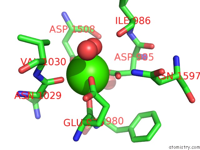
Mono view
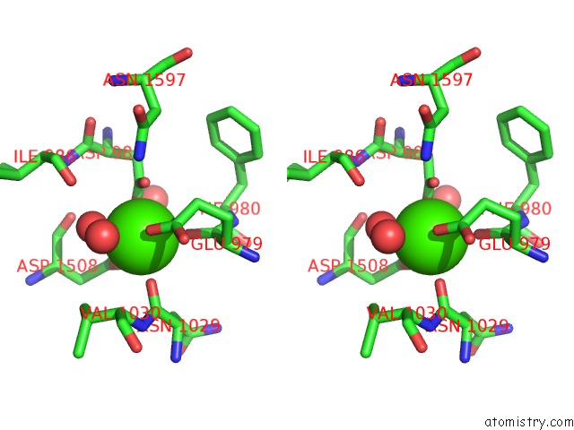
Stereo pair view
Mono view
Stereo pair view
A full contact list of Calcium with other atoms in the Ca binding
site number 1 of Crystal Structure of Lactobacillus Reuteri N-Terminally Truncated Glucansucrase GTF180 in Triclinic Apo- Form within 5.0Å range:
|
Reference:
A.Vujicic-Zagar,
T.Pijning,
S.Kralj,
C.A.Lopez,
W.Eeuwema,
L.Dijkhuizen,
B.W.Dijkstra.
Crystal Structure of A 117 kDa Glucansucrase Fragment Provides Insight Into Evolution and Product Specificity of GH70 Enzymes Proc.Natl.Acad.Sci.Usa V. 107 21406 2010.
ISSN: ISSN 0027-8424
PubMed: 21118988
DOI: 10.1073/PNAS.1007531107
Page generated: Sat Jul 13 12:18:53 2024
ISSN: ISSN 0027-8424
PubMed: 21118988
DOI: 10.1073/PNAS.1007531107
Last articles
Zn in 9J0NZn in 9J0O
Zn in 9J0P
Zn in 9FJX
Zn in 9EKB
Zn in 9C0F
Zn in 9CAH
Zn in 9CH0
Zn in 9CH3
Zn in 9CH1