Calcium »
PDB 3l7f-3ljt »
3lhm »
Calcium in PDB 3lhm: Crystal Structures of the Apo-and Holomutant Human Lysozymes with An Introduced CA2+ Binding Site
Enzymatic activity of Crystal Structures of the Apo-and Holomutant Human Lysozymes with An Introduced CA2+ Binding Site
All present enzymatic activity of Crystal Structures of the Apo-and Holomutant Human Lysozymes with An Introduced CA2+ Binding Site:
3.2.1.17;
3.2.1.17;
Protein crystallography data
The structure of Crystal Structures of the Apo-and Holomutant Human Lysozymes with An Introduced CA2+ Binding Site, PDB code: 3lhm
was solved by
K.Inaka,
M.Matsushima,
with X-Ray Crystallography technique. A brief refinement statistics is given in the table below:
| Resolution Low / High (Å) | N/A / 1.80 |
| Space group | P 21 21 21 |
| Cell size a, b, c (Å), α, β, γ (°) | 57.390, 60.970, 33.690, 90.00, 90.00, 90.00 |
| R / Rfree (%) | n/a / n/a |
Calcium Binding Sites:
The binding sites of Calcium atom in the Crystal Structures of the Apo-and Holomutant Human Lysozymes with An Introduced CA2+ Binding Site
(pdb code 3lhm). This binding sites where shown within
5.0 Angstroms radius around Calcium atom.
In total only one binding site of Calcium was determined in the Crystal Structures of the Apo-and Holomutant Human Lysozymes with An Introduced CA2+ Binding Site, PDB code: 3lhm:
In total only one binding site of Calcium was determined in the Crystal Structures of the Apo-and Holomutant Human Lysozymes with An Introduced CA2+ Binding Site, PDB code: 3lhm:
Calcium binding site 1 out of 1 in 3lhm
Go back to
Calcium binding site 1 out
of 1 in the Crystal Structures of the Apo-and Holomutant Human Lysozymes with An Introduced CA2+ Binding Site
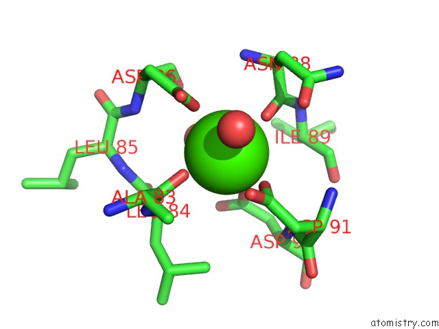
Mono view
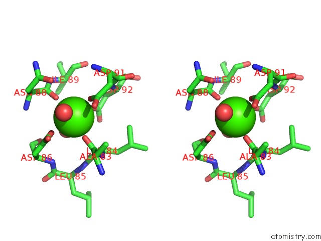
Stereo pair view
Mono view
Stereo pair view
A full contact list of Calcium with other atoms in the Ca binding
site number 1 of Crystal Structures of the Apo-and Holomutant Human Lysozymes with An Introduced CA2+ Binding Site within 5.0Å range:
|
Reference:
K.Inaka,
R.Kuroki,
M.Kikuchi,
M.Matsushima.
Crystal Structures of the Apo- and Holomutant Human Lysozymes with An Introduced CA2+ Binding Site. J.Biol.Chem. V. 266 20666 1991.
ISSN: ISSN 0021-9258
PubMed: 1939116
Page generated: Sat Jul 13 13:02:16 2024
ISSN: ISSN 0021-9258
PubMed: 1939116
Last articles
Zn in 9J0NZn in 9J0O
Zn in 9J0P
Zn in 9FJX
Zn in 9EKB
Zn in 9C0F
Zn in 9CAH
Zn in 9CH0
Zn in 9CH3
Zn in 9CH1