Calcium »
PDB 3m0c-3mip »
3m8d »
Calcium in PDB 3m8d: Crystal Structure of Spin-Labeled Btub V10R1 with Bound Calcium and Cyanocobalamin
Protein crystallography data
The structure of Crystal Structure of Spin-Labeled Btub V10R1 with Bound Calcium and Cyanocobalamin, PDB code: 3m8d
was solved by
D.M.Freed,
P.S.Horanyi,
M.C.Wiener,
D.S.Cafiso,
with X-Ray Crystallography technique. A brief refinement statistics is given in the table below:
| Resolution Low / High (Å) | 44.04 / 2.44 |
| Space group | P 31 2 1 |
| Cell size a, b, c (Å), α, β, γ (°) | 82.051, 82.051, 224.457, 90.00, 90.00, 120.00 |
| R / Rfree (%) | 22.6 / 27.5 |
Other elements in 3m8d:
The structure of Crystal Structure of Spin-Labeled Btub V10R1 with Bound Calcium and Cyanocobalamin also contains other interesting chemical elements:
| Cobalt | (Co) | 1 atom |
Calcium Binding Sites:
The binding sites of Calcium atom in the Crystal Structure of Spin-Labeled Btub V10R1 with Bound Calcium and Cyanocobalamin
(pdb code 3m8d). This binding sites where shown within
5.0 Angstroms radius around Calcium atom.
In total 3 binding sites of Calcium where determined in the Crystal Structure of Spin-Labeled Btub V10R1 with Bound Calcium and Cyanocobalamin, PDB code: 3m8d:
Jump to Calcium binding site number: 1; 2; 3;
In total 3 binding sites of Calcium where determined in the Crystal Structure of Spin-Labeled Btub V10R1 with Bound Calcium and Cyanocobalamin, PDB code: 3m8d:
Jump to Calcium binding site number: 1; 2; 3;
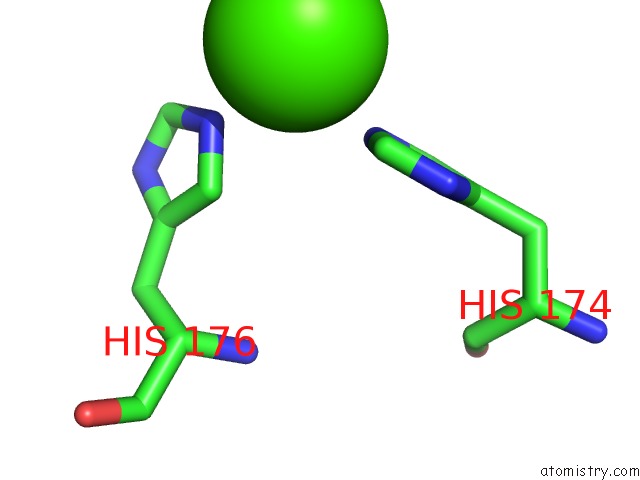
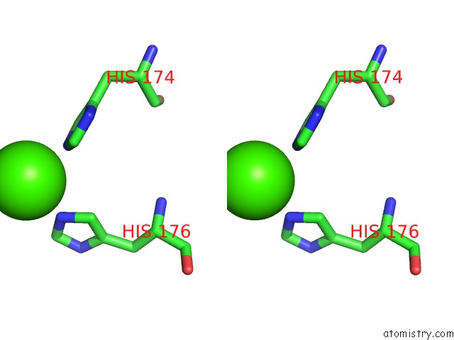
Calcium binding site 1 out of 3 in 3m8d
Go back to
Calcium binding site 1 out
of 3 in the Crystal Structure of Spin-Labeled Btub V10R1 with Bound Calcium and Cyanocobalamin
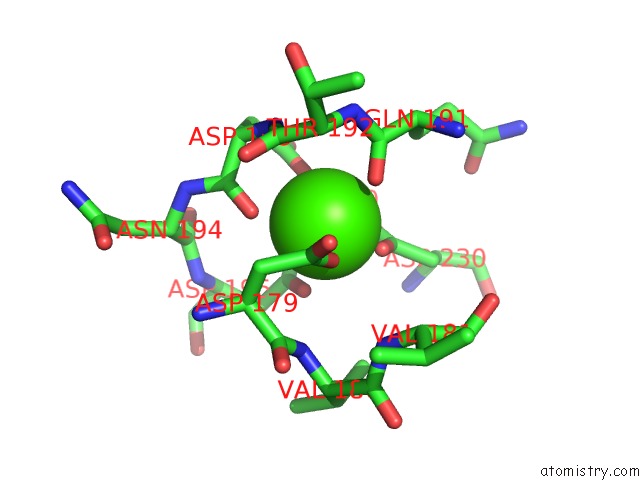
Mono view
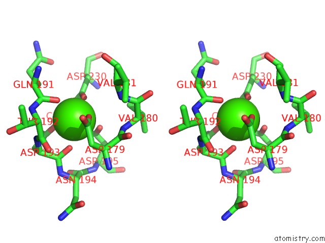
Stereo pair view
Mono view
Stereo pair view
A full contact list of Calcium with other atoms in the Ca binding
site number 1 of Crystal Structure of Spin-Labeled Btub V10R1 with Bound Calcium and Cyanocobalamin within 5.0Å range:
|
Calcium binding site 2 out of 3 in 3m8d
Go back to
Calcium binding site 2 out
of 3 in the Crystal Structure of Spin-Labeled Btub V10R1 with Bound Calcium and Cyanocobalamin
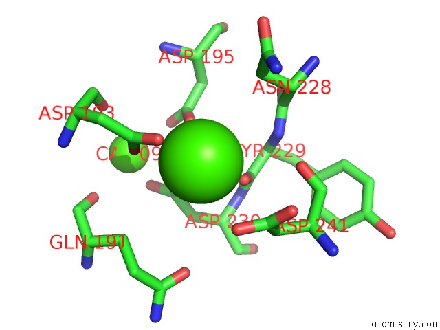
Mono view
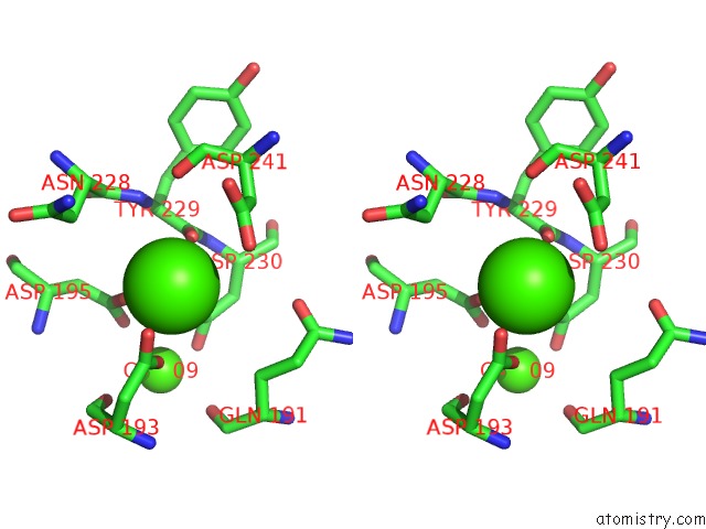
Stereo pair view
Mono view
Stereo pair view
A full contact list of Calcium with other atoms in the Ca binding
site number 2 of Crystal Structure of Spin-Labeled Btub V10R1 with Bound Calcium and Cyanocobalamin within 5.0Å range:
|
Calcium binding site 3 out of 3 in 3m8d
Go back to
Calcium binding site 3 out
of 3 in the Crystal Structure of Spin-Labeled Btub V10R1 with Bound Calcium and Cyanocobalamin
Mono view
Stereo pair view
Mono view
Stereo pair view
A full contact list of Calcium with other atoms in the Ca binding
site number 3 of Crystal Structure of Spin-Labeled Btub V10R1 with Bound Calcium and Cyanocobalamin within 5.0Å range:
|
Reference:
D.M.Freed,
P.S.Horanyi,
M.C.Wiener,
D.S.Cafiso.
Conformational Exchange in A Membrane Transport Protein Is Altered in Protein Crystals. Biophys.J. V. 99 1604 2010.
ISSN: ISSN 0006-3495
PubMed: 20816073
DOI: 10.1016/J.BPJ.2010.06.026
Page generated: Tue Jul 8 14:32:35 2025
ISSN: ISSN 0006-3495
PubMed: 20816073
DOI: 10.1016/J.BPJ.2010.06.026
Last articles
F in 4HT0F in 4HNA
F in 4HPX
F in 4HQH
F in 4HNS
F in 4HPJ
F in 4HN4
F in 4HJX
F in 4HLH
F in 4HL4