Calcium »
PDB 3m0c-3mip »
3mbr »
Calcium in PDB 3mbr: Crystal Structure of the Glutaminyl Cyclase From Xanthomonas Campestris
Enzymatic activity of Crystal Structure of the Glutaminyl Cyclase From Xanthomonas Campestris
All present enzymatic activity of Crystal Structure of the Glutaminyl Cyclase From Xanthomonas Campestris:
2.3.2.5;
2.3.2.5;
Protein crystallography data
The structure of Crystal Structure of the Glutaminyl Cyclase From Xanthomonas Campestris, PDB code: 3mbr
was solved by
W.-L.Huang,
Y.-R.Wang,
T.-P.Ko,
C.-Y.Chia,
K.-F.Huang,
A.H.-J.Wang,
with X-Ray Crystallography technique. A brief refinement statistics is given in the table below:
| Resolution Low / High (Å) | 29.31 / 1.44 |
| Space group | I 4 |
| Cell size a, b, c (Å), α, β, γ (°) | 95.330, 95.330, 65.088, 90.00, 90.00, 90.00 |
| R / Rfree (%) | 17.4 / 20.9 |
Calcium Binding Sites:
The binding sites of Calcium atom in the Crystal Structure of the Glutaminyl Cyclase From Xanthomonas Campestris
(pdb code 3mbr). This binding sites where shown within
5.0 Angstroms radius around Calcium atom.
In total only one binding site of Calcium was determined in the Crystal Structure of the Glutaminyl Cyclase From Xanthomonas Campestris, PDB code: 3mbr:
In total only one binding site of Calcium was determined in the Crystal Structure of the Glutaminyl Cyclase From Xanthomonas Campestris, PDB code: 3mbr:
Calcium binding site 1 out of 1 in 3mbr
Go back to
Calcium binding site 1 out
of 1 in the Crystal Structure of the Glutaminyl Cyclase From Xanthomonas Campestris
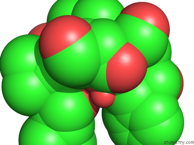
Mono view
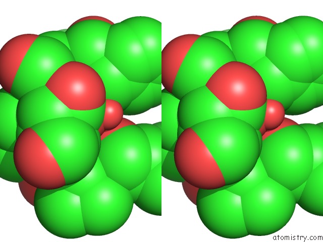
Stereo pair view
Mono view
Stereo pair view
A full contact list of Calcium with other atoms in the Ca binding
site number 1 of Crystal Structure of the Glutaminyl Cyclase From Xanthomonas Campestris within 5.0Å range:
|
Reference:
W.-L.Huang,
Y.-R.Wang,
T.-P.Ko,
C.-Y.Chia,
K.-F.Huang,
A.H.-J.Wang.
Crystal Structure and Functional Analysis of the Glutaminyl Cyclase From Xanthomonas Campestris J.Mol.Biol. V. 401 374 2010.
ISSN: ISSN 0022-2836
PubMed: 20558177
DOI: 10.1016/J.JMB.2010.06.012
Page generated: Sat Jul 13 13:34:02 2024
ISSN: ISSN 0022-2836
PubMed: 20558177
DOI: 10.1016/J.JMB.2010.06.012
Last articles
Zn in 9J0NZn in 9J0O
Zn in 9J0P
Zn in 9FJX
Zn in 9EKB
Zn in 9C0F
Zn in 9CAH
Zn in 9CH0
Zn in 9CH3
Zn in 9CH1