Calcium »
PDB 3m0c-3mip »
3mcy »
Calcium in PDB 3mcy: Crystal Structure of Fimh Lectin Domain Bound to Biphenyl Mannoside Meta-Methyl Ester.
Protein crystallography data
The structure of Crystal Structure of Fimh Lectin Domain Bound to Biphenyl Mannoside Meta-Methyl Ester., PDB code: 3mcy
was solved by
B.A.Ford,
S.J.Hultgren,
with X-Ray Crystallography technique. A brief refinement statistics is given in the table below:
| Resolution Low / High (Å) | 59.95 / 2.90 |
| Space group | P 41 3 2 |
| Cell size a, b, c (Å), α, β, γ (°) | 198.820, 198.820, 198.820, 90.00, 90.00, 90.00 |
| R / Rfree (%) | 21.9 / 24.5 |
Other elements in 3mcy:
The structure of Crystal Structure of Fimh Lectin Domain Bound to Biphenyl Mannoside Meta-Methyl Ester. also contains other interesting chemical elements:
| Chlorine | (Cl) | 3 atoms |
Calcium Binding Sites:
The binding sites of Calcium atom in the Crystal Structure of Fimh Lectin Domain Bound to Biphenyl Mannoside Meta-Methyl Ester.
(pdb code 3mcy). This binding sites where shown within
5.0 Angstroms radius around Calcium atom.
In total 4 binding sites of Calcium where determined in the Crystal Structure of Fimh Lectin Domain Bound to Biphenyl Mannoside Meta-Methyl Ester., PDB code: 3mcy:
Jump to Calcium binding site number: 1; 2; 3; 4;
In total 4 binding sites of Calcium where determined in the Crystal Structure of Fimh Lectin Domain Bound to Biphenyl Mannoside Meta-Methyl Ester., PDB code: 3mcy:
Jump to Calcium binding site number: 1; 2; 3; 4;
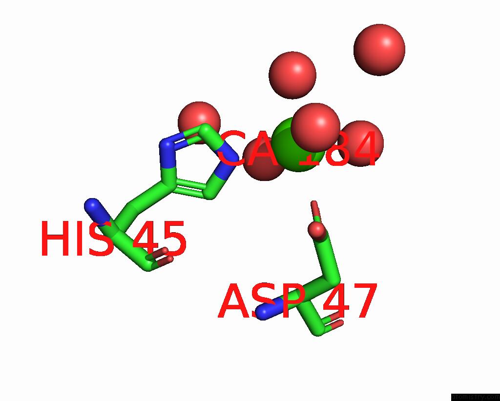
Calcium binding site 1 out of 4 in 3mcy
Go back to
Calcium binding site 1 out
of 4 in the Crystal Structure of Fimh Lectin Domain Bound to Biphenyl Mannoside Meta-Methyl Ester.
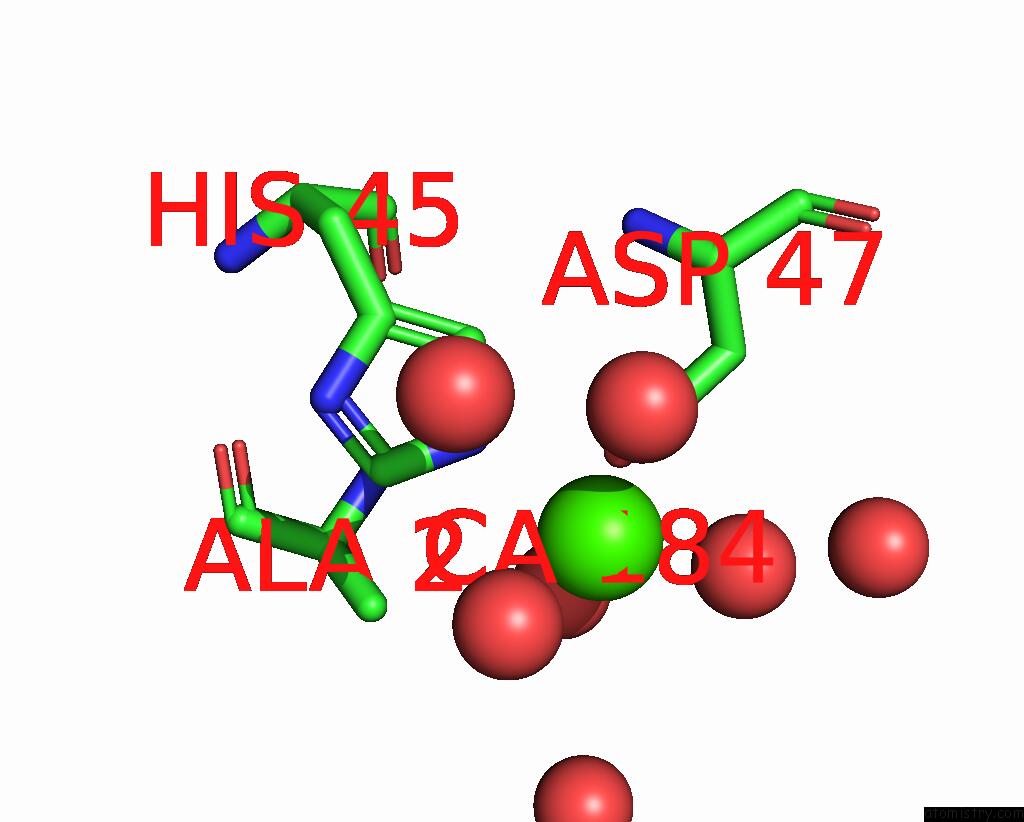
Mono view
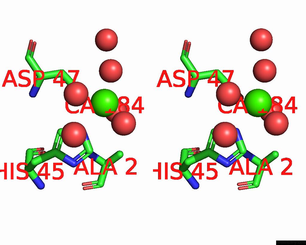
Stereo pair view
Mono view
Stereo pair view
A full contact list of Calcium with other atoms in the Ca binding
site number 1 of Crystal Structure of Fimh Lectin Domain Bound to Biphenyl Mannoside Meta-Methyl Ester. within 5.0Å range:
|
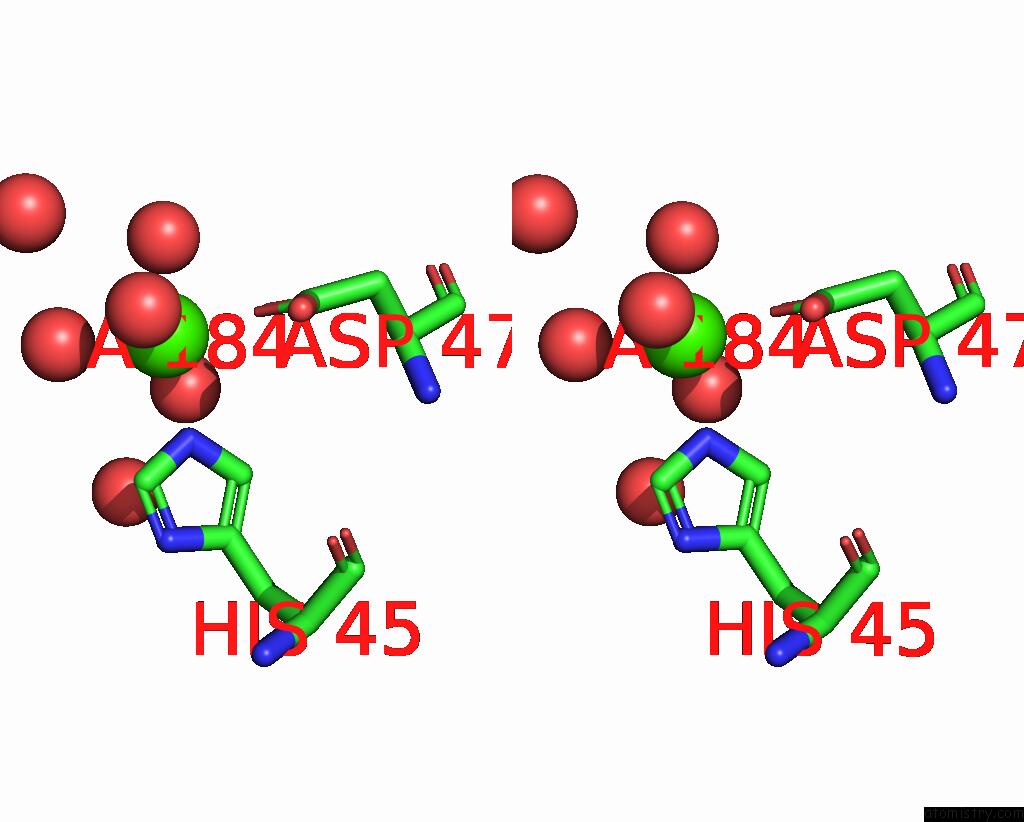
Calcium binding site 2 out of 4 in 3mcy
Go back to
Calcium binding site 2 out
of 4 in the Crystal Structure of Fimh Lectin Domain Bound to Biphenyl Mannoside Meta-Methyl Ester.
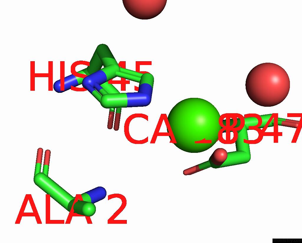
Mono view
Stereo pair view
Mono view
Stereo pair view
A full contact list of Calcium with other atoms in the Ca binding
site number 2 of Crystal Structure of Fimh Lectin Domain Bound to Biphenyl Mannoside Meta-Methyl Ester. within 5.0Å range:
|
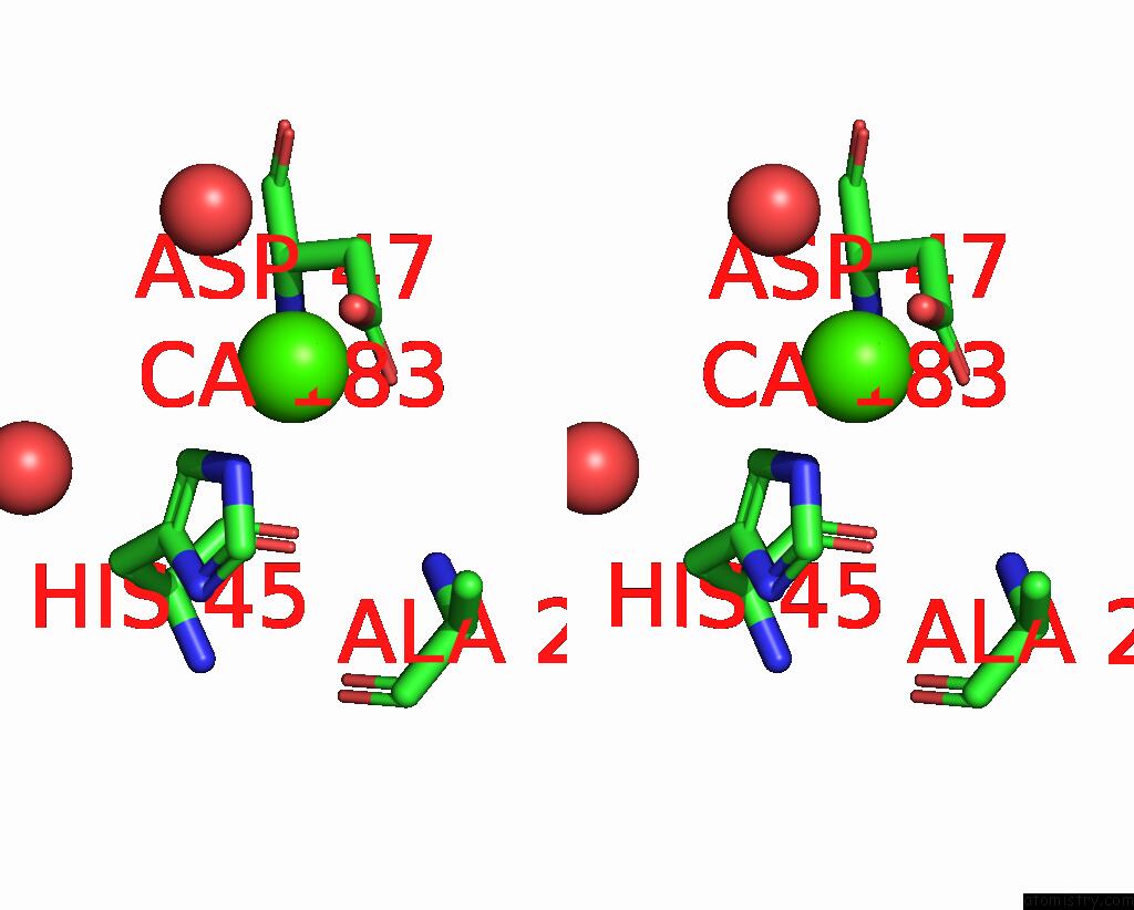
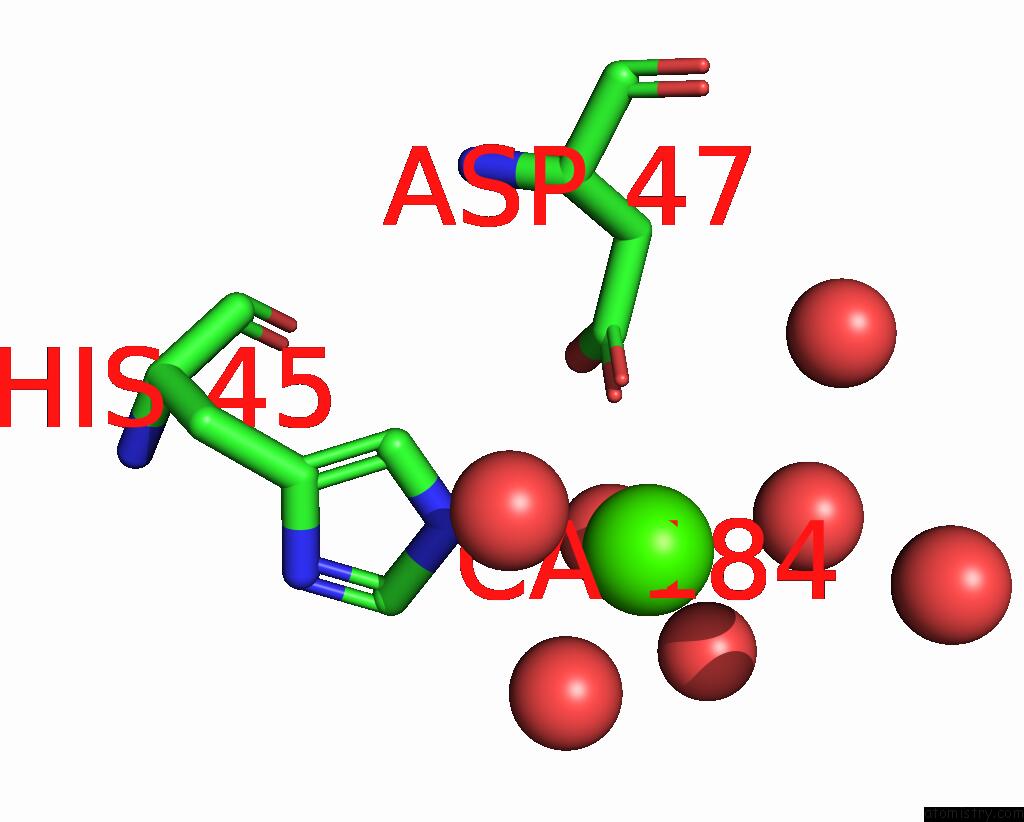
Calcium binding site 3 out of 4 in 3mcy
Go back to
Calcium binding site 3 out
of 4 in the Crystal Structure of Fimh Lectin Domain Bound to Biphenyl Mannoside Meta-Methyl Ester.
Mono view
Stereo pair view
Mono view
Stereo pair view
A full contact list of Calcium with other atoms in the Ca binding
site number 3 of Crystal Structure of Fimh Lectin Domain Bound to Biphenyl Mannoside Meta-Methyl Ester. within 5.0Å range:
|
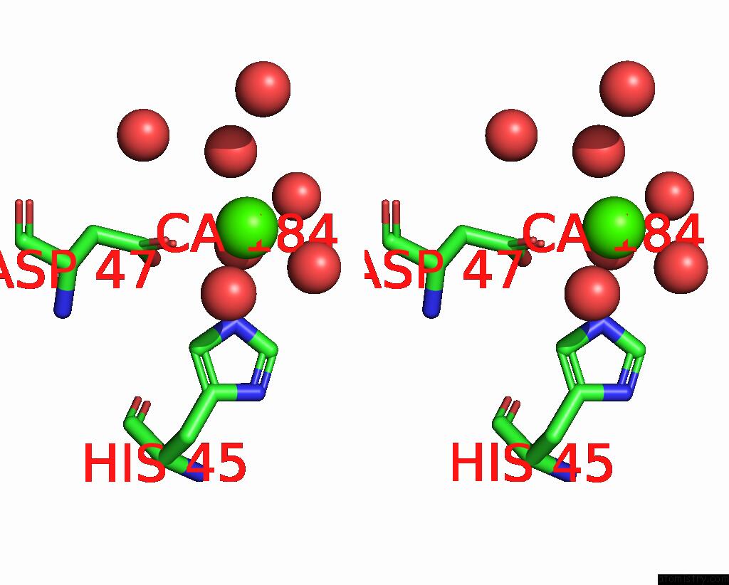
Calcium binding site 4 out of 4 in 3mcy
Go back to
Calcium binding site 4 out
of 4 in the Crystal Structure of Fimh Lectin Domain Bound to Biphenyl Mannoside Meta-Methyl Ester.
Mono view
Stereo pair view
Mono view
Stereo pair view
A full contact list of Calcium with other atoms in the Ca binding
site number 4 of Crystal Structure of Fimh Lectin Domain Bound to Biphenyl Mannoside Meta-Methyl Ester. within 5.0Å range:
|
Reference:
Z.Han,
J.S.Pinkner,
B.Ford,
R.Obermann,
W.Nolan,
S.A.Wildman,
D.Hobbs,
T.Ellenberger,
C.K.Cusumano,
S.J.Hultgren,
J.W.Janetka.
Structure-Based Drug Design and Optimization of Mannoside Bacterial Fimh Antagonists. J.Med.Chem. V. 53 4779 2010.
ISSN: ISSN 0022-2623
PubMed: 20507142
DOI: 10.1021/JM100438S
Page generated: Sat Jul 13 13:34:18 2024
ISSN: ISSN 0022-2623
PubMed: 20507142
DOI: 10.1021/JM100438S
Last articles
Zn in 9J0NZn in 9J0O
Zn in 9J0P
Zn in 9FJX
Zn in 9EKB
Zn in 9C0F
Zn in 9CAH
Zn in 9CH0
Zn in 9CH3
Zn in 9CH1