Calcium »
PDB 3m0c-3mip »
3mdo »
Calcium in PDB 3mdo: Crystal Structure of A Putative Phosphoribosylformylglycinamidine Cyclo-Ligase (BDI_2101) From Parabacteroides Distasonis Atcc 8503 at 1.91 A Resolution
Protein crystallography data
The structure of Crystal Structure of A Putative Phosphoribosylformylglycinamidine Cyclo-Ligase (BDI_2101) From Parabacteroides Distasonis Atcc 8503 at 1.91 A Resolution, PDB code: 3mdo
was solved by
Joint Center For Structural Genomics (Jcsg),
with X-Ray Crystallography technique. A brief refinement statistics is given in the table below:
| Resolution Low / High (Å) | 29.62 / 1.91 |
| Space group | P 41 21 2 |
| Cell size a, b, c (Å), α, β, γ (°) | 99.945, 99.945, 163.020, 90.00, 90.00, 90.00 |
| R / Rfree (%) | 15.4 / 19 |
Other elements in 3mdo:
The structure of Crystal Structure of A Putative Phosphoribosylformylglycinamidine Cyclo-Ligase (BDI_2101) From Parabacteroides Distasonis Atcc 8503 at 1.91 A Resolution also contains other interesting chemical elements:
| Arsenic | (As) | 1 atom |
| Chlorine | (Cl) | 4 atoms |
Calcium Binding Sites:
The binding sites of Calcium atom in the Crystal Structure of A Putative Phosphoribosylformylglycinamidine Cyclo-Ligase (BDI_2101) From Parabacteroides Distasonis Atcc 8503 at 1.91 A Resolution
(pdb code 3mdo). This binding sites where shown within
5.0 Angstroms radius around Calcium atom.
In total 7 binding sites of Calcium where determined in the Crystal Structure of A Putative Phosphoribosylformylglycinamidine Cyclo-Ligase (BDI_2101) From Parabacteroides Distasonis Atcc 8503 at 1.91 A Resolution, PDB code: 3mdo:
Jump to Calcium binding site number: 1; 2; 3; 4; 5; 6; 7;
In total 7 binding sites of Calcium where determined in the Crystal Structure of A Putative Phosphoribosylformylglycinamidine Cyclo-Ligase (BDI_2101) From Parabacteroides Distasonis Atcc 8503 at 1.91 A Resolution, PDB code: 3mdo:
Jump to Calcium binding site number: 1; 2; 3; 4; 5; 6; 7;
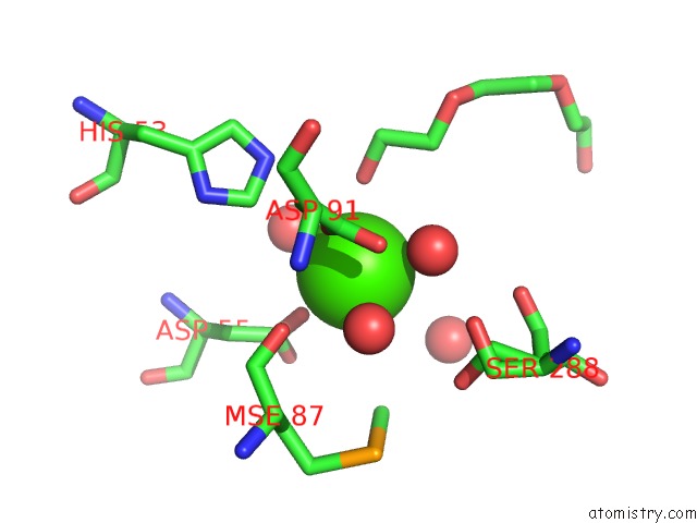
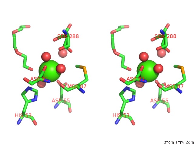
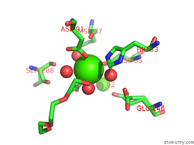
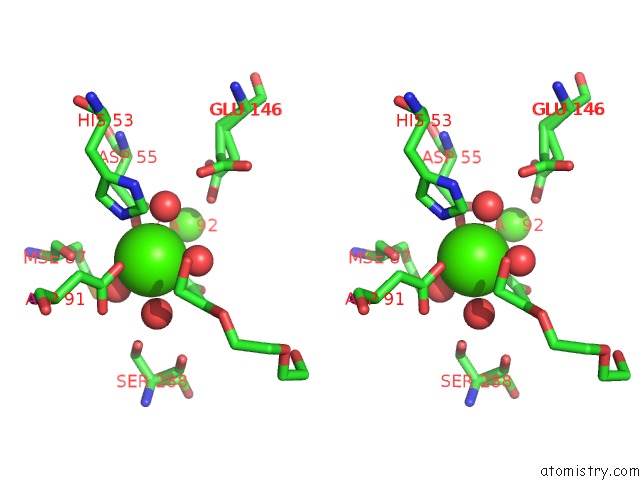
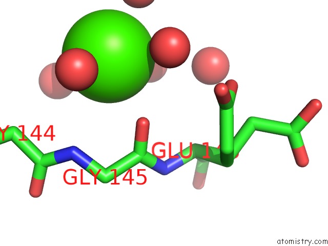
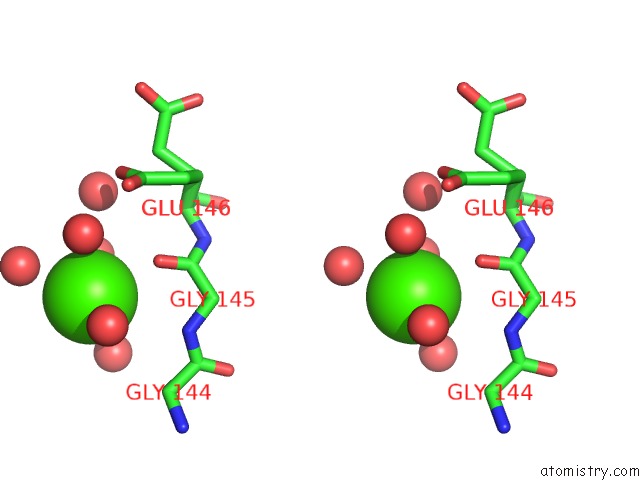
Calcium binding site 1 out of 7 in 3mdo
Go back to
Calcium binding site 1 out
of 7 in the Crystal Structure of A Putative Phosphoribosylformylglycinamidine Cyclo-Ligase (BDI_2101) From Parabacteroides Distasonis Atcc 8503 at 1.91 A Resolution
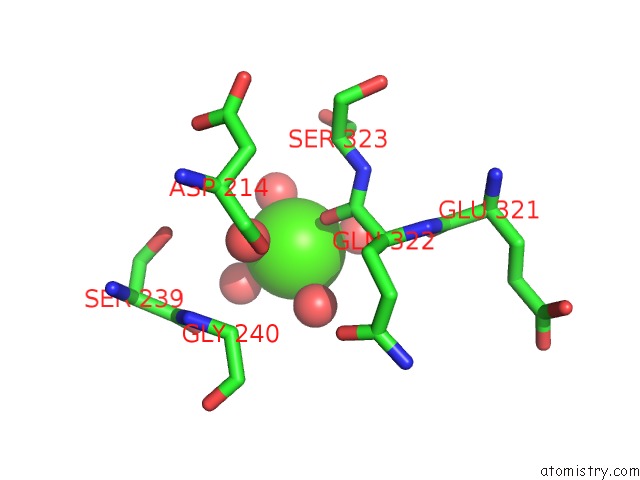
Mono view
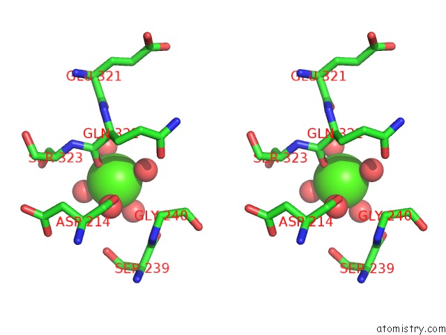
Stereo pair view
Mono view
Stereo pair view
A full contact list of Calcium with other atoms in the Ca binding
site number 1 of Crystal Structure of A Putative Phosphoribosylformylglycinamidine Cyclo-Ligase (BDI_2101) From Parabacteroides Distasonis Atcc 8503 at 1.91 A Resolution within 5.0Å range:
|
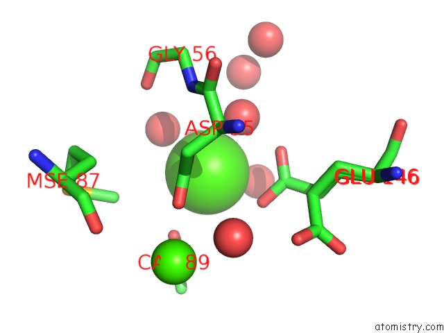
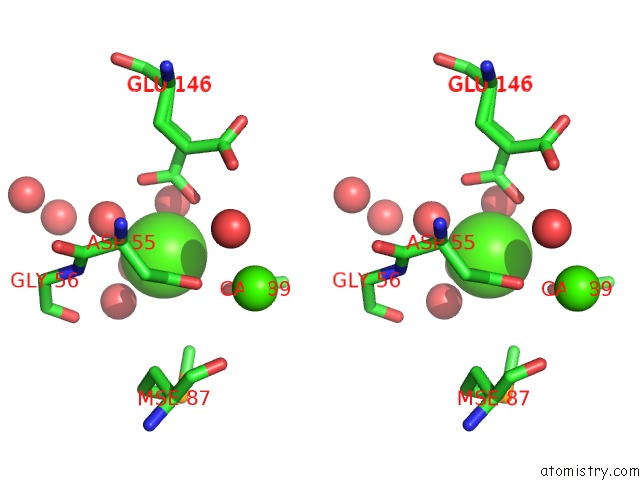
Calcium binding site 2 out of 7 in 3mdo
Go back to
Calcium binding site 2 out
of 7 in the Crystal Structure of A Putative Phosphoribosylformylglycinamidine Cyclo-Ligase (BDI_2101) From Parabacteroides Distasonis Atcc 8503 at 1.91 A Resolution
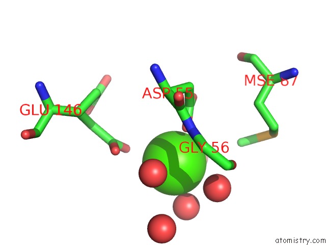
Mono view
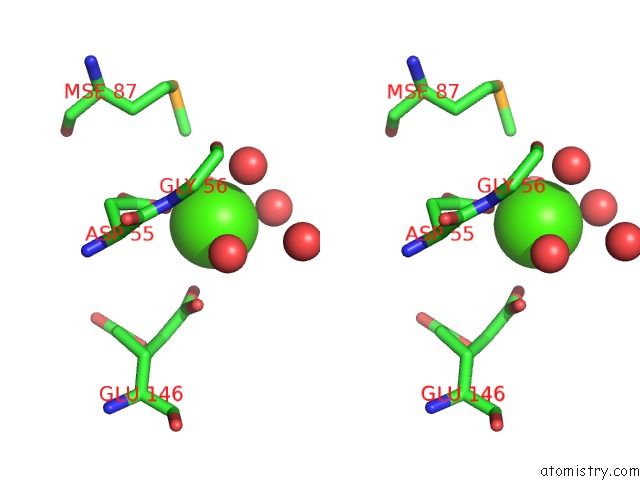
Stereo pair view
Mono view
Stereo pair view
A full contact list of Calcium with other atoms in the Ca binding
site number 2 of Crystal Structure of A Putative Phosphoribosylformylglycinamidine Cyclo-Ligase (BDI_2101) From Parabacteroides Distasonis Atcc 8503 at 1.91 A Resolution within 5.0Å range:
|
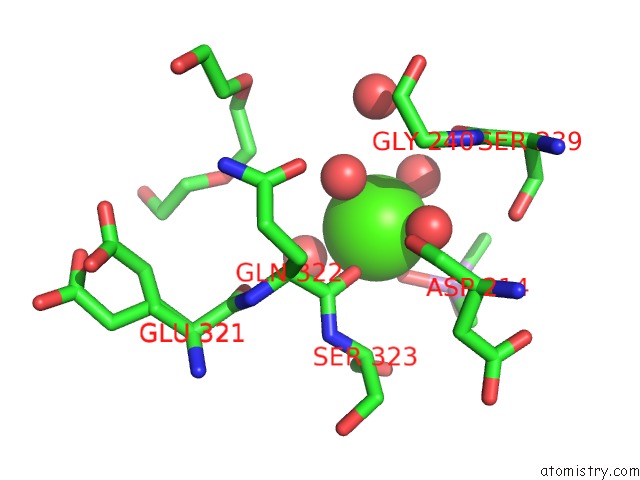
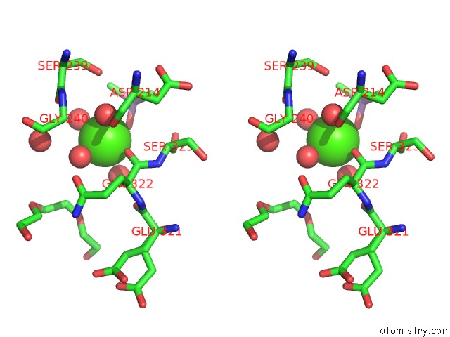
Calcium binding site 3 out of 7 in 3mdo
Go back to
Calcium binding site 3 out
of 7 in the Crystal Structure of A Putative Phosphoribosylformylglycinamidine Cyclo-Ligase (BDI_2101) From Parabacteroides Distasonis Atcc 8503 at 1.91 A Resolution
Mono view
Stereo pair view
Mono view
Stereo pair view
A full contact list of Calcium with other atoms in the Ca binding
site number 3 of Crystal Structure of A Putative Phosphoribosylformylglycinamidine Cyclo-Ligase (BDI_2101) From Parabacteroides Distasonis Atcc 8503 at 1.91 A Resolution within 5.0Å range:
|
Calcium binding site 4 out of 7 in 3mdo
Go back to
Calcium binding site 4 out
of 7 in the Crystal Structure of A Putative Phosphoribosylformylglycinamidine Cyclo-Ligase (BDI_2101) From Parabacteroides Distasonis Atcc 8503 at 1.91 A Resolution
Mono view
Stereo pair view
Mono view
Stereo pair view
A full contact list of Calcium with other atoms in the Ca binding
site number 4 of Crystal Structure of A Putative Phosphoribosylformylglycinamidine Cyclo-Ligase (BDI_2101) From Parabacteroides Distasonis Atcc 8503 at 1.91 A Resolution within 5.0Å range:
|
Calcium binding site 5 out of 7 in 3mdo
Go back to
Calcium binding site 5 out
of 7 in the Crystal Structure of A Putative Phosphoribosylformylglycinamidine Cyclo-Ligase (BDI_2101) From Parabacteroides Distasonis Atcc 8503 at 1.91 A Resolution
Mono view
Stereo pair view
Mono view
Stereo pair view
A full contact list of Calcium with other atoms in the Ca binding
site number 5 of Crystal Structure of A Putative Phosphoribosylformylglycinamidine Cyclo-Ligase (BDI_2101) From Parabacteroides Distasonis Atcc 8503 at 1.91 A Resolution within 5.0Å range:
|
Calcium binding site 6 out of 7 in 3mdo
Go back to
Calcium binding site 6 out
of 7 in the Crystal Structure of A Putative Phosphoribosylformylglycinamidine Cyclo-Ligase (BDI_2101) From Parabacteroides Distasonis Atcc 8503 at 1.91 A Resolution
Mono view
Stereo pair view
Mono view
Stereo pair view
A full contact list of Calcium with other atoms in the Ca binding
site number 6 of Crystal Structure of A Putative Phosphoribosylformylglycinamidine Cyclo-Ligase (BDI_2101) From Parabacteroides Distasonis Atcc 8503 at 1.91 A Resolution within 5.0Å range:
|
Calcium binding site 7 out of 7 in 3mdo
Go back to
Calcium binding site 7 out
of 7 in the Crystal Structure of A Putative Phosphoribosylformylglycinamidine Cyclo-Ligase (BDI_2101) From Parabacteroides Distasonis Atcc 8503 at 1.91 A Resolution
Mono view
Stereo pair view
Mono view
Stereo pair view
A full contact list of Calcium with other atoms in the Ca binding
site number 7 of Crystal Structure of A Putative Phosphoribosylformylglycinamidine Cyclo-Ligase (BDI_2101) From Parabacteroides Distasonis Atcc 8503 at 1.91 A Resolution within 5.0Å range:
|
Reference:
Joint Center For Structural Genomics (Jcsg),
Joint Center For Structural Genomics (Jcsg).
N/A N/A.
Page generated: Sat Jul 13 13:34:37 2024
Last articles
Zn in 9J0NZn in 9J0O
Zn in 9J0P
Zn in 9FJX
Zn in 9EKB
Zn in 9C0F
Zn in 9CAH
Zn in 9CH0
Zn in 9CH3
Zn in 9CH1