Calcium »
PDB 3mis-3mz5 »
3msa »
Calcium in PDB 3msa: Crystal Structure of Thermolysin in Complex with 3-Bromophenol
Enzymatic activity of Crystal Structure of Thermolysin in Complex with 3-Bromophenol
All present enzymatic activity of Crystal Structure of Thermolysin in Complex with 3-Bromophenol:
3.4.24.27;
3.4.24.27;
Protein crystallography data
The structure of Crystal Structure of Thermolysin in Complex with 3-Bromophenol, PDB code: 3msa
was solved by
J.Behnen,
A.Heine,
G.Klebe,
with X-Ray Crystallography technique. A brief refinement statistics is given in the table below:
| Resolution Low / High (Å) | 10.00 / 1.66 |
| Space group | P 61 2 2 |
| Cell size a, b, c (Å), α, β, γ (°) | 93.111, 93.111, 128.726, 90.00, 90.00, 120.00 |
| R / Rfree (%) | 16.6 / 20 |
Other elements in 3msa:
The structure of Crystal Structure of Thermolysin in Complex with 3-Bromophenol also contains other interesting chemical elements:
| Bromine | (Br) | 2 atoms |
| Zinc | (Zn) | 1 atom |
Calcium Binding Sites:
The binding sites of Calcium atom in the Crystal Structure of Thermolysin in Complex with 3-Bromophenol
(pdb code 3msa). This binding sites where shown within
5.0 Angstroms radius around Calcium atom.
In total 4 binding sites of Calcium where determined in the Crystal Structure of Thermolysin in Complex with 3-Bromophenol, PDB code: 3msa:
Jump to Calcium binding site number: 1; 2; 3; 4;
In total 4 binding sites of Calcium where determined in the Crystal Structure of Thermolysin in Complex with 3-Bromophenol, PDB code: 3msa:
Jump to Calcium binding site number: 1; 2; 3; 4;
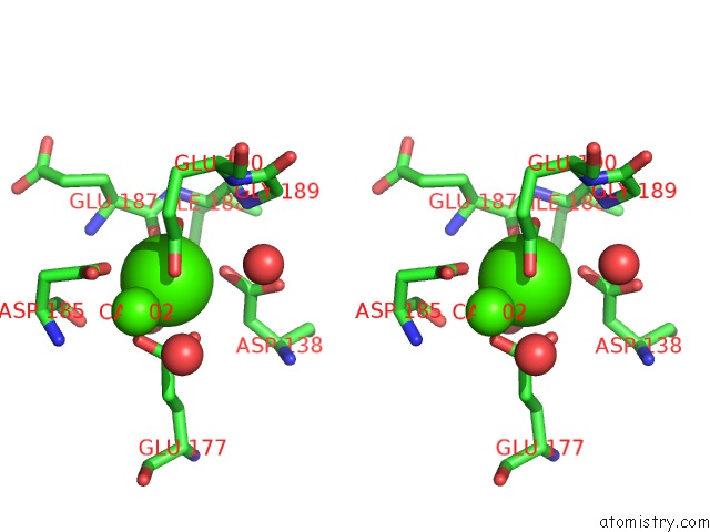
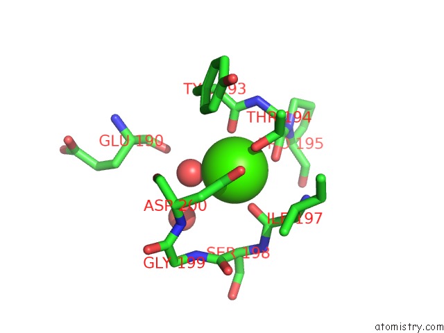
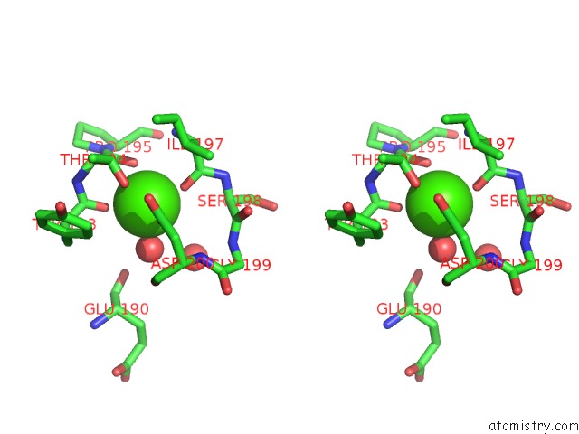
Calcium binding site 1 out of 4 in 3msa
Go back to
Calcium binding site 1 out
of 4 in the Crystal Structure of Thermolysin in Complex with 3-Bromophenol

Mono view
Stereo pair view
Mono view
Stereo pair view
A full contact list of Calcium with other atoms in the Ca binding
site number 1 of Crystal Structure of Thermolysin in Complex with 3-Bromophenol within 5.0Å range:
|
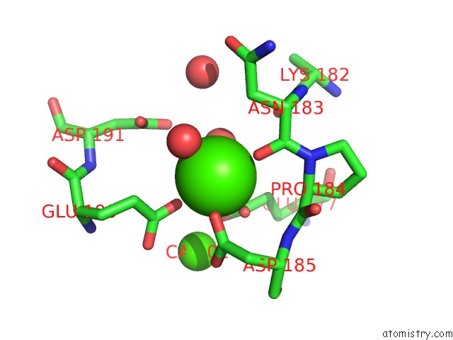
Calcium binding site 2 out of 4 in 3msa
Go back to
Calcium binding site 2 out
of 4 in the Crystal Structure of Thermolysin in Complex with 3-Bromophenol
Mono view
Stereo pair view
Mono view
Stereo pair view
A full contact list of Calcium with other atoms in the Ca binding
site number 2 of Crystal Structure of Thermolysin in Complex with 3-Bromophenol within 5.0Å range:
|
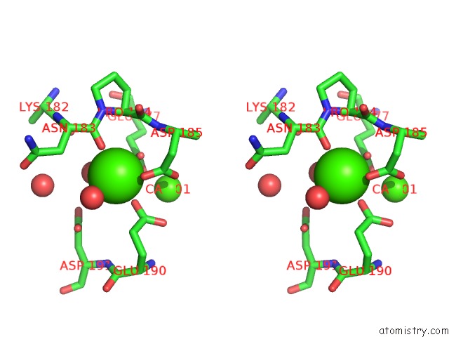
Calcium binding site 3 out of 4 in 3msa
Go back to
Calcium binding site 3 out
of 4 in the Crystal Structure of Thermolysin in Complex with 3-Bromophenol

Mono view
Stereo pair view
Mono view
Stereo pair view
A full contact list of Calcium with other atoms in the Ca binding
site number 3 of Crystal Structure of Thermolysin in Complex with 3-Bromophenol within 5.0Å range:
|
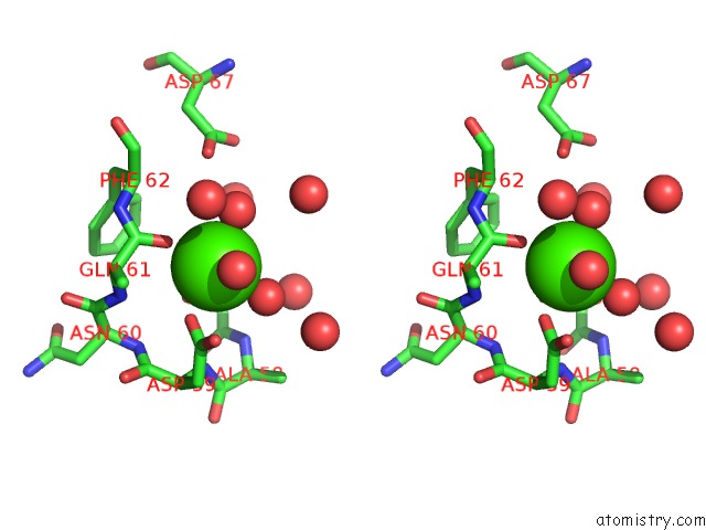
Calcium binding site 4 out of 4 in 3msa
Go back to
Calcium binding site 4 out
of 4 in the Crystal Structure of Thermolysin in Complex with 3-Bromophenol
Mono view
Stereo pair view
Mono view
Stereo pair view
A full contact list of Calcium with other atoms in the Ca binding
site number 4 of Crystal Structure of Thermolysin in Complex with 3-Bromophenol within 5.0Å range:
|
Reference:
J.Behnen,
H.Koster,
G.Neudert,
T.Craan,
A.Heine,
G.Klebe.
Experimental and Computational Active Site Mapping As A Starting Point to Fragment-Based Lead Discovery. Chemmedchem V. 7 248 2012.
ISSN: ISSN 1860-7179
PubMed: 22213702
DOI: 10.1002/CMDC.201100490
Page generated: Sat Jul 13 13:43:31 2024
ISSN: ISSN 1860-7179
PubMed: 22213702
DOI: 10.1002/CMDC.201100490
Last articles
Zn in 9MJ5Zn in 9HNW
Zn in 9G0L
Zn in 9FNE
Zn in 9DZN
Zn in 9E0I
Zn in 9D32
Zn in 9DAK
Zn in 8ZXC
Zn in 8ZUF