Calcium »
PDB 3mis-3mz5 »
3mvs »
Calcium in PDB 3mvs: Structure of the N-Terminus of Cadherin 23
Protein crystallography data
The structure of Structure of the N-Terminus of Cadherin 23, PDB code: 3mvs
was solved by
P.Clark,
J.S.Joseph,
A.R.Kolatkar,
with X-Ray Crystallography technique. A brief refinement statistics is given in the table below:
| Resolution Low / High (Å) | 23.00 / 1.10 |
| Space group | P 1 21 1 |
| Cell size a, b, c (Å), α, β, γ (°) | 38.375, 64.208, 47.859, 90.00, 110.89, 90.00 |
| R / Rfree (%) | 16.3 / 18.8 |
Calcium Binding Sites:
The binding sites of Calcium atom in the Structure of the N-Terminus of Cadherin 23
(pdb code 3mvs). This binding sites where shown within
5.0 Angstroms radius around Calcium atom.
In total 6 binding sites of Calcium where determined in the Structure of the N-Terminus of Cadherin 23, PDB code: 3mvs:
Jump to Calcium binding site number: 1; 2; 3; 4; 5; 6;
In total 6 binding sites of Calcium where determined in the Structure of the N-Terminus of Cadherin 23, PDB code: 3mvs:
Jump to Calcium binding site number: 1; 2; 3; 4; 5; 6;
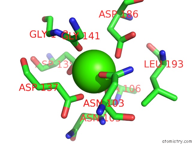
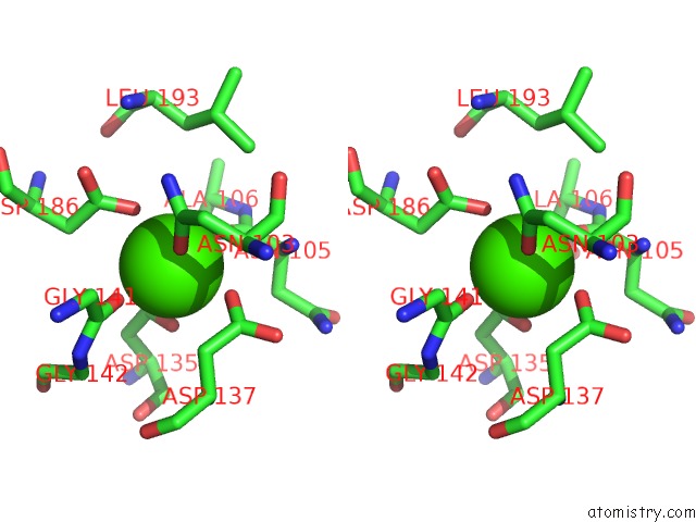
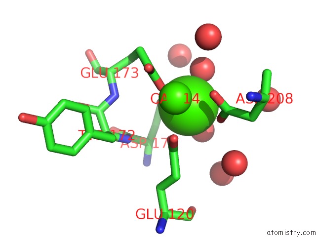
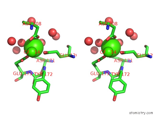
Calcium binding site 1 out of 6 in 3mvs
Go back to
Calcium binding site 1 out
of 6 in the Structure of the N-Terminus of Cadherin 23
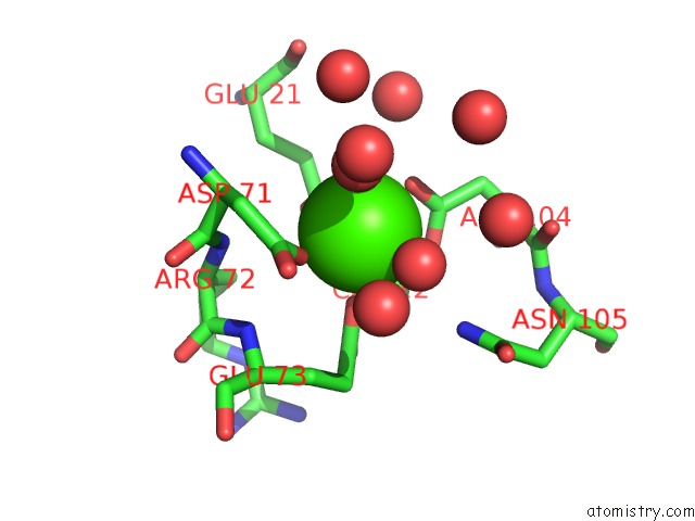
Mono view
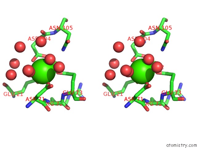
Stereo pair view
Mono view
Stereo pair view
A full contact list of Calcium with other atoms in the Ca binding
site number 1 of Structure of the N-Terminus of Cadherin 23 within 5.0Å range:
|
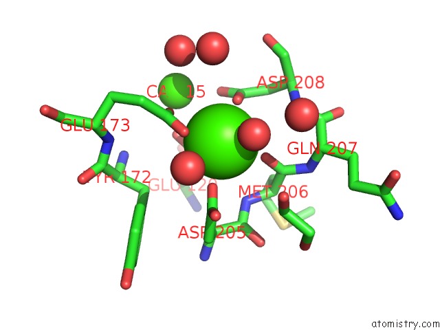
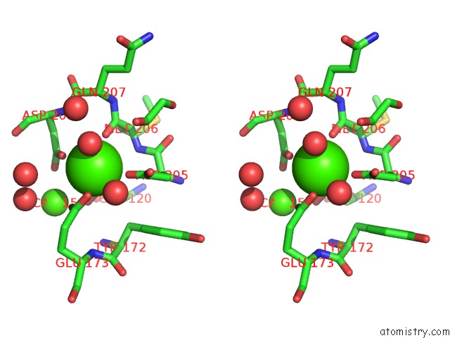
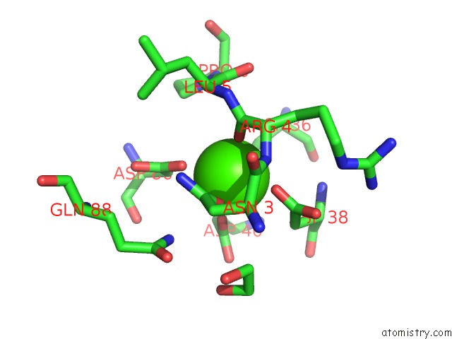
Calcium binding site 2 out of 6 in 3mvs
Go back to
Calcium binding site 2 out
of 6 in the Structure of the N-Terminus of Cadherin 23
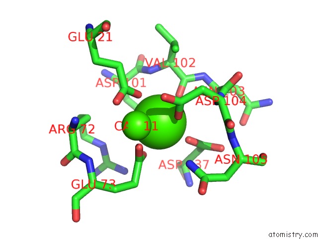
Mono view
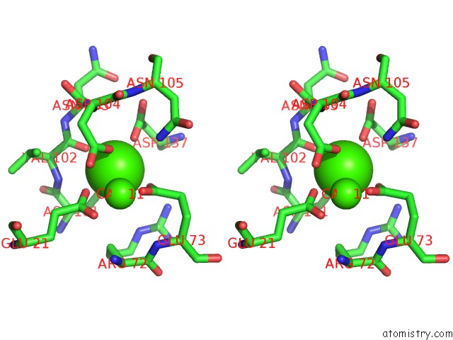
Stereo pair view
Mono view
Stereo pair view
A full contact list of Calcium with other atoms in the Ca binding
site number 2 of Structure of the N-Terminus of Cadherin 23 within 5.0Å range:
|
Calcium binding site 3 out of 6 in 3mvs
Go back to
Calcium binding site 3 out
of 6 in the Structure of the N-Terminus of Cadherin 23
Mono view
Stereo pair view
Mono view
Stereo pair view
A full contact list of Calcium with other atoms in the Ca binding
site number 3 of Structure of the N-Terminus of Cadherin 23 within 5.0Å range:
|
Calcium binding site 4 out of 6 in 3mvs
Go back to
Calcium binding site 4 out
of 6 in the Structure of the N-Terminus of Cadherin 23
Mono view
Stereo pair view
Mono view
Stereo pair view
A full contact list of Calcium with other atoms in the Ca binding
site number 4 of Structure of the N-Terminus of Cadherin 23 within 5.0Å range:
|
Calcium binding site 5 out of 6 in 3mvs
Go back to
Calcium binding site 5 out
of 6 in the Structure of the N-Terminus of Cadherin 23
Mono view
Stereo pair view
Mono view
Stereo pair view
A full contact list of Calcium with other atoms in the Ca binding
site number 5 of Structure of the N-Terminus of Cadherin 23 within 5.0Å range:
|
Calcium binding site 6 out of 6 in 3mvs
Go back to
Calcium binding site 6 out
of 6 in the Structure of the N-Terminus of Cadherin 23
Mono view

Stereo pair view
Mono view
Stereo pair view
A full contact list of Calcium with other atoms in the Ca binding
site number 6 of Structure of the N-Terminus of Cadherin 23 within 5.0Å range:
|
Reference:
H.M.Elledge,
P.Kazmierczak,
P.Clark,
J.S.Joseph,
A.Kolatkar,
P.Kuhn,
U.Muller.
Structure of the N Terminus of Cadherin 23 Reveals A New Adhesion Mechanism For A Subset of Cadherin Superfamily Members. Proc.Natl.Acad.Sci.Usa V. 107 10708 2010.
ISSN: ISSN 0027-8424
PubMed: 20498078
DOI: 10.1073/PNAS.1006284107
Page generated: Sat Jul 13 13:45:19 2024
ISSN: ISSN 0027-8424
PubMed: 20498078
DOI: 10.1073/PNAS.1006284107
Last articles
Zn in 9J0NZn in 9J0O
Zn in 9J0P
Zn in 9FJX
Zn in 9EKB
Zn in 9C0F
Zn in 9CAH
Zn in 9CH0
Zn in 9CH3
Zn in 9CH1