Calcium »
PDB 3mzo-3ngi »
3n8g »
Calcium in PDB 3n8g: Structure of the (Sr)CA2+-Atpase CA2-E1-Caamppcp Form
Enzymatic activity of Structure of the (Sr)CA2+-Atpase CA2-E1-Caamppcp Form
All present enzymatic activity of Structure of the (Sr)CA2+-Atpase CA2-E1-Caamppcp Form:
3.6.3.8;
3.6.3.8;
Protein crystallography data
The structure of Structure of the (Sr)CA2+-Atpase CA2-E1-Caamppcp Form, PDB code: 3n8g
was solved by
M.Bublitz,
C.Olesen,
H.Poulsen,
J.P.Morth,
J.V.Moller,
P.Nissen,
with X-Ray Crystallography technique. A brief refinement statistics is given in the table below:
| Resolution Low / High (Å) | 30.13 / 2.58 |
| Space group | C 1 2 1 |
| Cell size a, b, c (Å), α, β, γ (°) | 162.000, 76.000, 151.000, 90.00, 108.00, 90.00 |
| R / Rfree (%) | 20.2 / 24.5 |
Other elements in 3n8g:
The structure of Structure of the (Sr)CA2+-Atpase CA2-E1-Caamppcp Form also contains other interesting chemical elements:
| Potassium | (K) | 1 atom |
Calcium Binding Sites:
The binding sites of Calcium atom in the Structure of the (Sr)CA2+-Atpase CA2-E1-Caamppcp Form
(pdb code 3n8g). This binding sites where shown within
5.0 Angstroms radius around Calcium atom.
In total 3 binding sites of Calcium where determined in the Structure of the (Sr)CA2+-Atpase CA2-E1-Caamppcp Form, PDB code: 3n8g:
Jump to Calcium binding site number: 1; 2; 3;
In total 3 binding sites of Calcium where determined in the Structure of the (Sr)CA2+-Atpase CA2-E1-Caamppcp Form, PDB code: 3n8g:
Jump to Calcium binding site number: 1; 2; 3;
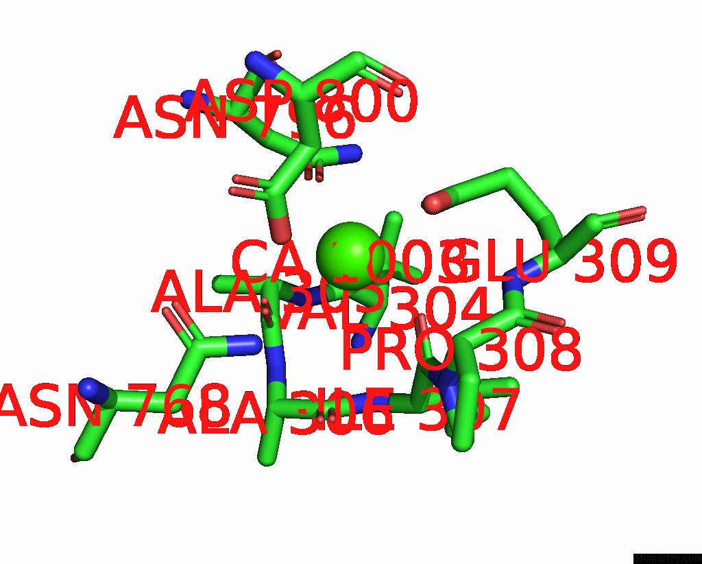
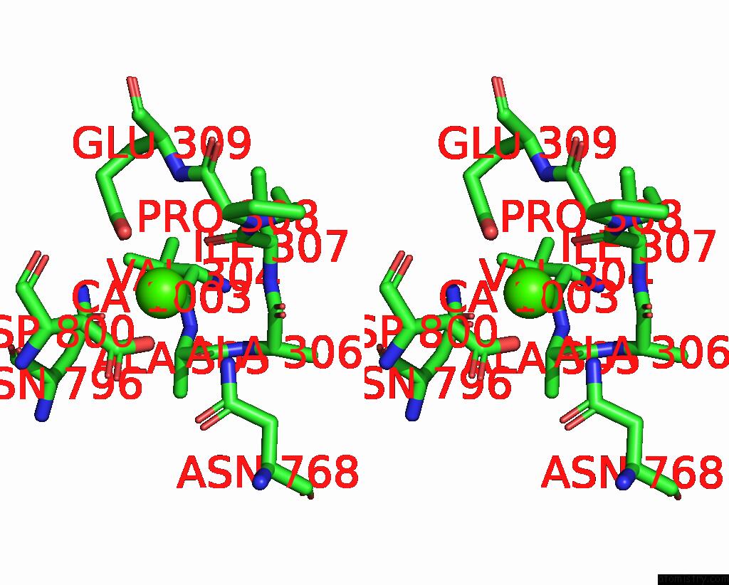
Calcium binding site 1 out of 3 in 3n8g
Go back to
Calcium binding site 1 out
of 3 in the Structure of the (Sr)CA2+-Atpase CA2-E1-Caamppcp Form

Mono view

Stereo pair view
Mono view
Stereo pair view
A full contact list of Calcium with other atoms in the Ca binding
site number 1 of Structure of the (Sr)CA2+-Atpase CA2-E1-Caamppcp Form within 5.0Å range:
|
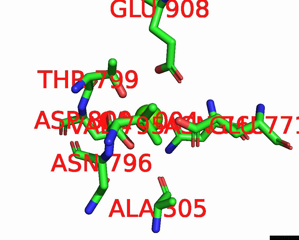
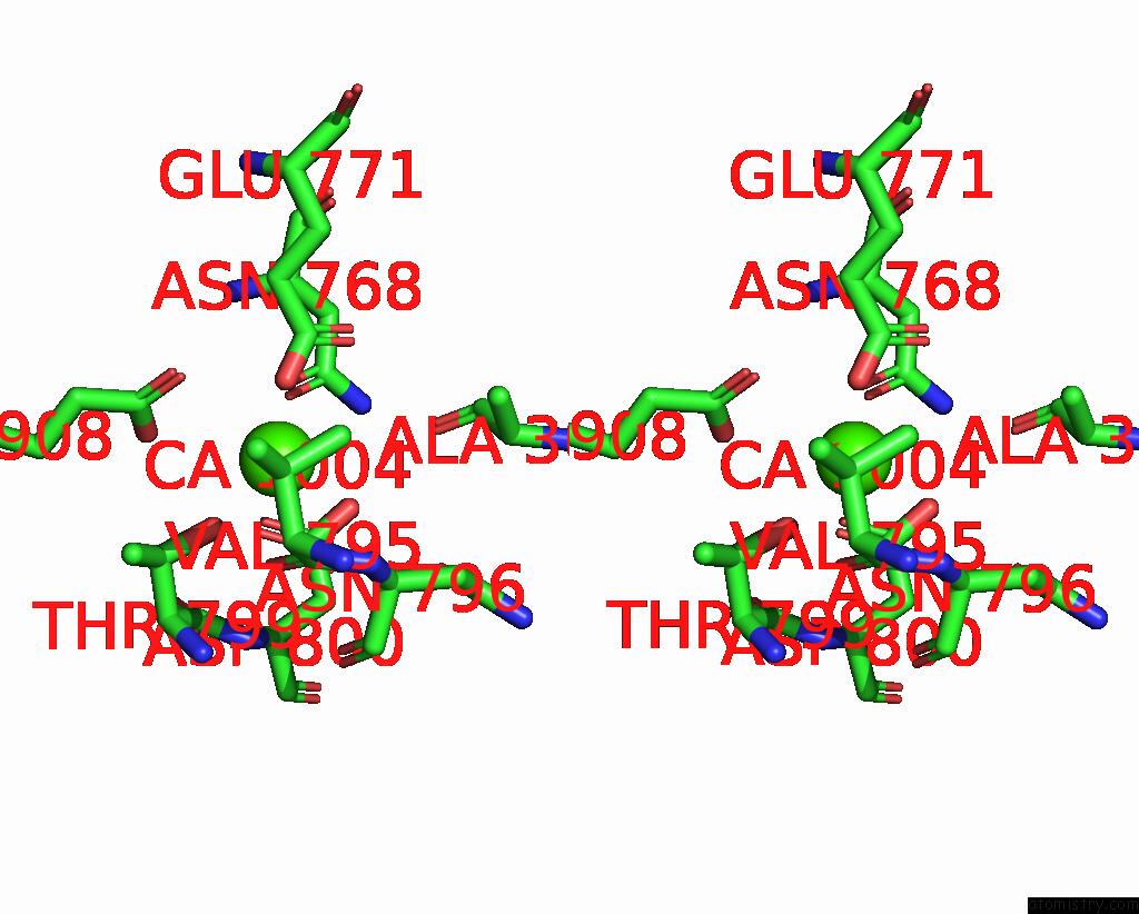
Calcium binding site 2 out of 3 in 3n8g
Go back to
Calcium binding site 2 out
of 3 in the Structure of the (Sr)CA2+-Atpase CA2-E1-Caamppcp Form
Mono view
Stereo pair view
Mono view
Stereo pair view
A full contact list of Calcium with other atoms in the Ca binding
site number 2 of Structure of the (Sr)CA2+-Atpase CA2-E1-Caamppcp Form within 5.0Å range:
|
Calcium binding site 3 out of 3 in 3n8g
Go back to
Calcium binding site 3 out
of 3 in the Structure of the (Sr)CA2+-Atpase CA2-E1-Caamppcp Form
Mono view
Stereo pair view
Mono view
Stereo pair view
A full contact list of Calcium with other atoms in the Ca binding
site number 3 of Structure of the (Sr)CA2+-Atpase CA2-E1-Caamppcp Form within 5.0Å range:
|
Reference:
M.Bublitz,
M.Musgaard,
H.Poulsen,
L.Thogersen,
C.Olesen,
B.Schiott,
J.P.Morth,
J.V.Moller,
P.Nissen.
Ion Pathways in the Sarcoplasmic Reticulum CA2+-Atpase. J.Biol.Chem. V. 288 10759 2013.
ISSN: ISSN 0021-9258
PubMed: 23400778
DOI: 10.1074/JBC.R112.436550
Page generated: Sat Jul 13 14:53:41 2024
ISSN: ISSN 0021-9258
PubMed: 23400778
DOI: 10.1074/JBC.R112.436550
Last articles
Zn in 9J0NZn in 9J0O
Zn in 9J0P
Zn in 9FJX
Zn in 9EKB
Zn in 9C0F
Zn in 9CAH
Zn in 9CH0
Zn in 9CH3
Zn in 9CH1