Calcium »
PDB 3mzo-3ngi »
3n9k »
Calcium in PDB 3n9k: F229A/E292S Double Mutant of Exo-Beta-1,3-Glucanase From Candida Albicans in Complex with Laminaritriose at 1.7 A
Enzymatic activity of F229A/E292S Double Mutant of Exo-Beta-1,3-Glucanase From Candida Albicans in Complex with Laminaritriose at 1.7 A
All present enzymatic activity of F229A/E292S Double Mutant of Exo-Beta-1,3-Glucanase From Candida Albicans in Complex with Laminaritriose at 1.7 A:
3.2.1.58;
3.2.1.58;
Protein crystallography data
The structure of F229A/E292S Double Mutant of Exo-Beta-1,3-Glucanase From Candida Albicans in Complex with Laminaritriose at 1.7 A, PDB code: 3n9k
was solved by
Y.Nakatani,
S.M.Cutfield,
J.F.Cutfield,
with X-Ray Crystallography technique. A brief refinement statistics is given in the table below:
| Resolution Low / High (Å) | 38.19 / 1.70 |
| Space group | P 21 21 21 |
| Cell size a, b, c (Å), α, β, γ (°) | 58.725, 64.397, 94.866, 90.00, 90.00, 90.00 |
| R / Rfree (%) | 15.2 / 19.7 |
Calcium Binding Sites:
The binding sites of Calcium atom in the F229A/E292S Double Mutant of Exo-Beta-1,3-Glucanase From Candida Albicans in Complex with Laminaritriose at 1.7 A
(pdb code 3n9k). This binding sites where shown within
5.0 Angstroms radius around Calcium atom.
In total only one binding site of Calcium was determined in the F229A/E292S Double Mutant of Exo-Beta-1,3-Glucanase From Candida Albicans in Complex with Laminaritriose at 1.7 A, PDB code: 3n9k:
In total only one binding site of Calcium was determined in the F229A/E292S Double Mutant of Exo-Beta-1,3-Glucanase From Candida Albicans in Complex with Laminaritriose at 1.7 A, PDB code: 3n9k:
Calcium binding site 1 out of 1 in 3n9k
Go back to
Calcium binding site 1 out
of 1 in the F229A/E292S Double Mutant of Exo-Beta-1,3-Glucanase From Candida Albicans in Complex with Laminaritriose at 1.7 A
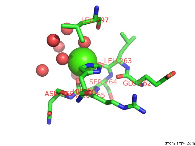
Mono view
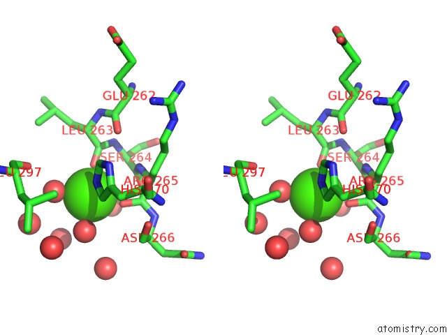
Stereo pair view
Mono view
Stereo pair view
A full contact list of Calcium with other atoms in the Ca binding
site number 1 of F229A/E292S Double Mutant of Exo-Beta-1,3-Glucanase From Candida Albicans in Complex with Laminaritriose at 1.7 A within 5.0Å range:
|
Reference:
W.M.Patrick,
Y.Nakatani,
S.M.Cutfield,
M.L.Sharpe,
R.J.Ramsay,
J.F.Cutfield.
Carbohydrate Binding Sites in Candida Albicans Exo-Beta-1,3-Glucanase and the Role of the Phe-Phe 'Clamp' at the Active Site Entrance Febs J. V. 277 4549 2010.
ISSN: ISSN 1742-464X
PubMed: 20875088
DOI: 10.1111/J.1742-4658.2010.07869.X
Page generated: Sat Jul 13 14:55:11 2024
ISSN: ISSN 1742-464X
PubMed: 20875088
DOI: 10.1111/J.1742-4658.2010.07869.X
Last articles
Zn in 9J0NZn in 9J0O
Zn in 9J0P
Zn in 9FJX
Zn in 9EKB
Zn in 9C0F
Zn in 9CAH
Zn in 9CH0
Zn in 9CH3
Zn in 9CH1