Calcium »
PDB 3ngw-3nuc »
3np6 »
Calcium in PDB 3np6: The Crystal Structure of Berberine Bound to Dna D(Cgtacg)
Protein crystallography data
The structure of The Crystal Structure of Berberine Bound to Dna D(Cgtacg), PDB code: 3np6
was solved by
M.Ferraroni,
C.Bazzicalupi,
P.Gratteri,
A.R.Bilia,
with X-Ray Crystallography technique. A brief refinement statistics is given in the table below:
| Resolution Low / High (Å) | 39.42 / 2.30 |
| Space group | P 32 2 1 |
| Cell size a, b, c (Å), α, β, γ (°) | 30.370, 30.370, 118.260, 90.00, 90.00, 120.00 |
| R / Rfree (%) | 23.6 / 30.1 |
Calcium Binding Sites:
The binding sites of Calcium atom in the The Crystal Structure of Berberine Bound to Dna D(Cgtacg)
(pdb code 3np6). This binding sites where shown within
5.0 Angstroms radius around Calcium atom.
In total only one binding site of Calcium was determined in the The Crystal Structure of Berberine Bound to Dna D(Cgtacg), PDB code: 3np6:
In total only one binding site of Calcium was determined in the The Crystal Structure of Berberine Bound to Dna D(Cgtacg), PDB code: 3np6:
Calcium binding site 1 out of 1 in 3np6
Go back to
Calcium binding site 1 out
of 1 in the The Crystal Structure of Berberine Bound to Dna D(Cgtacg)
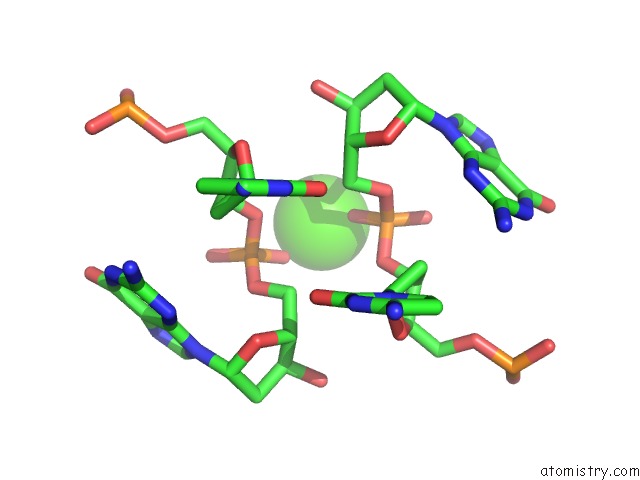
Mono view
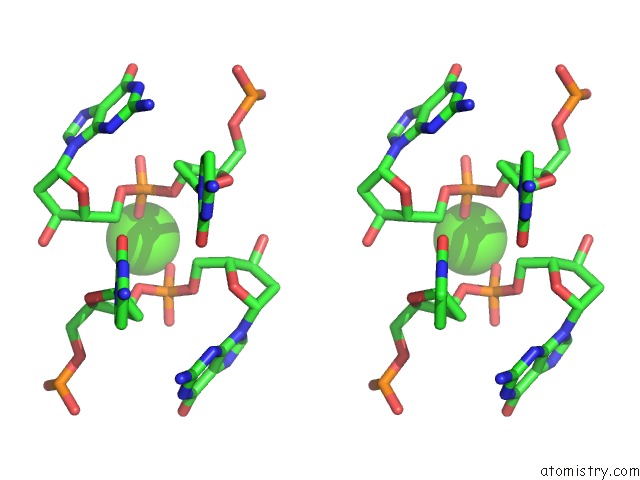
Stereo pair view
Mono view
Stereo pair view
A full contact list of Calcium with other atoms in the Ca binding
site number 1 of The Crystal Structure of Berberine Bound to Dna D(Cgtacg) within 5.0Å range:
|
Reference:
M.Ferraroni,
C.Bazzicalupi,
A.R.Bilia,
P.Gratteri.
X-Ray Diffraction Analyses of the Natural Isoquinoline Alkaloids Berberine and Sanguinarine Complexed with Double Helix Dna D(Cgtacg) Chem.Commun.(Camb.) V. 47 4917 2011.
ISSN: ISSN 1359-7345
PubMed: 21431128
DOI: 10.1039/C1CC10971E
Page generated: Tue Jul 8 14:58:21 2025
ISSN: ISSN 1359-7345
PubMed: 21431128
DOI: 10.1039/C1CC10971E
Last articles
Cl in 8D5HCl in 8D4Z
Cl in 8D5B
Cl in 8D4I
Cl in 8D59
Cl in 8D4P
Cl in 8D3S
Cl in 8D1X
Cl in 8D3N
Cl in 8D3H