Calcium »
PDB 3nur-3ojn »
3o03 »
Calcium in PDB 3o03: Quaternary Complex Structure of Gluconate 5-Dehydrogenase From Streptococcus Suis Type 2
Enzymatic activity of Quaternary Complex Structure of Gluconate 5-Dehydrogenase From Streptococcus Suis Type 2
All present enzymatic activity of Quaternary Complex Structure of Gluconate 5-Dehydrogenase From Streptococcus Suis Type 2:
1.1.1.69;
1.1.1.69;
Protein crystallography data
The structure of Quaternary Complex Structure of Gluconate 5-Dehydrogenase From Streptococcus Suis Type 2, PDB code: 3o03
was solved by
H.Peng,
F.Gao,
Q.Zhang,
Y.Liu,
G.F.Gao,
with X-Ray Crystallography technique. A brief refinement statistics is given in the table below:
| Resolution Low / High (Å) | 25.87 / 1.90 |
| Space group | I 2 2 2 |
| Cell size a, b, c (Å), α, β, γ (°) | 71.244, 75.272, 98.700, 90.00, 90.00, 90.00 |
| R / Rfree (%) | 19.1 / 24.2 |
Calcium Binding Sites:
The binding sites of Calcium atom in the Quaternary Complex Structure of Gluconate 5-Dehydrogenase From Streptococcus Suis Type 2
(pdb code 3o03). This binding sites where shown within
5.0 Angstroms radius around Calcium atom.
In total only one binding site of Calcium was determined in the Quaternary Complex Structure of Gluconate 5-Dehydrogenase From Streptococcus Suis Type 2, PDB code: 3o03:
In total only one binding site of Calcium was determined in the Quaternary Complex Structure of Gluconate 5-Dehydrogenase From Streptococcus Suis Type 2, PDB code: 3o03:
Calcium binding site 1 out of 1 in 3o03
Go back to
Calcium binding site 1 out
of 1 in the Quaternary Complex Structure of Gluconate 5-Dehydrogenase From Streptococcus Suis Type 2
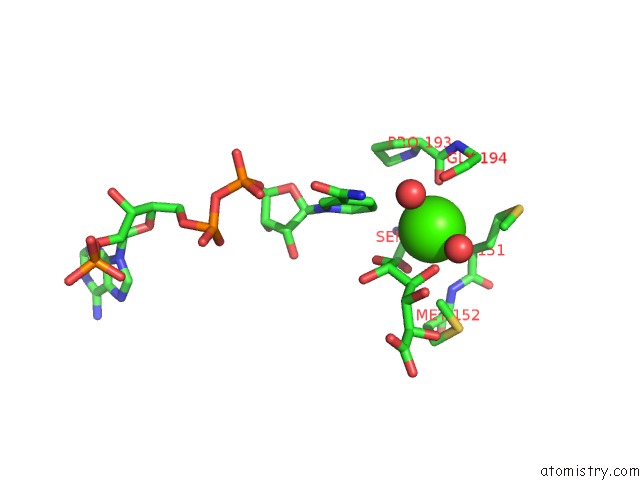
Mono view
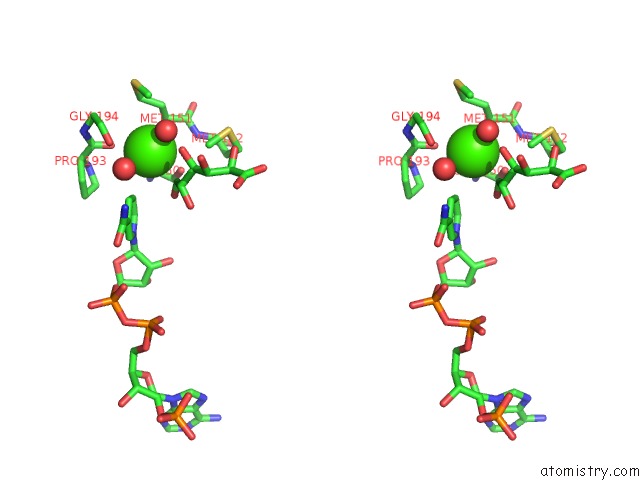
Stereo pair view
Mono view
Stereo pair view
A full contact list of Calcium with other atoms in the Ca binding
site number 1 of Quaternary Complex Structure of Gluconate 5-Dehydrogenase From Streptococcus Suis Type 2 within 5.0Å range:
|
Reference:
Q.Zhang,
H.Peng,
F.Gao,
Y.Liu,
H.Cheng,
J.Thompson,
G.F.Gao.
Structural Insight Into the Catalytic Mechanism of Gluconate 5-Dehydrogenase From Streptococcus Suis: Crystal Structures of the Substrate-Free and Quaternary Complex Enzymes. Protein Sci. V. 18 294 2009.
ISSN: ISSN 0961-8368
PubMed: 19177572
DOI: 10.1002/PRO.32
Page generated: Sat Jul 13 15:11:16 2024
ISSN: ISSN 0961-8368
PubMed: 19177572
DOI: 10.1002/PRO.32
Last articles
Zn in 9J0NZn in 9J0O
Zn in 9J0P
Zn in 9FJX
Zn in 9EKB
Zn in 9C0F
Zn in 9CAH
Zn in 9CH0
Zn in 9CH3
Zn in 9CH1