Calcium »
PDB 3nur-3ojn »
3o4p »
Calcium in PDB 3o4p: Dfpase at 0.85 Angstrom Resolution (H Atoms Included)
Enzymatic activity of Dfpase at 0.85 Angstrom Resolution (H Atoms Included)
All present enzymatic activity of Dfpase at 0.85 Angstrom Resolution (H Atoms Included):
3.1.8.2;
3.1.8.2;
Protein crystallography data
The structure of Dfpase at 0.85 Angstrom Resolution (H Atoms Included), PDB code: 3o4p
was solved by
D.Liebschner,
M.Elias,
J.Koepke,
C.Lecomte,
B.Guillot,
C.Jelsch,
E.Chabriere,
with X-Ray Crystallography technique. A brief refinement statistics is given in the table below:
| Resolution Low / High (Å) | 20.80 / 0.85 |
| Space group | P 21 21 21 |
| Cell size a, b, c (Å), α, β, γ (°) | 43.114, 81.849, 86.467, 90.00, 90.00, 90.00 |
| R / Rfree (%) | 10.3 / 12.1 |
Calcium Binding Sites:
The binding sites of Calcium atom in the Dfpase at 0.85 Angstrom Resolution (H Atoms Included)
(pdb code 3o4p). This binding sites where shown within
5.0 Angstroms radius around Calcium atom.
In total 2 binding sites of Calcium where determined in the Dfpase at 0.85 Angstrom Resolution (H Atoms Included), PDB code: 3o4p:
Jump to Calcium binding site number: 1; 2;
In total 2 binding sites of Calcium where determined in the Dfpase at 0.85 Angstrom Resolution (H Atoms Included), PDB code: 3o4p:
Jump to Calcium binding site number: 1; 2;
Calcium binding site 1 out of 2 in 3o4p
Go back to
Calcium binding site 1 out
of 2 in the Dfpase at 0.85 Angstrom Resolution (H Atoms Included)
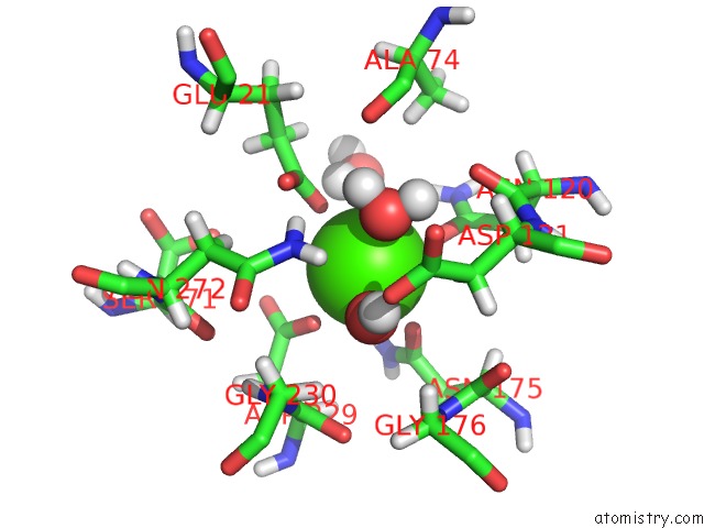
Mono view
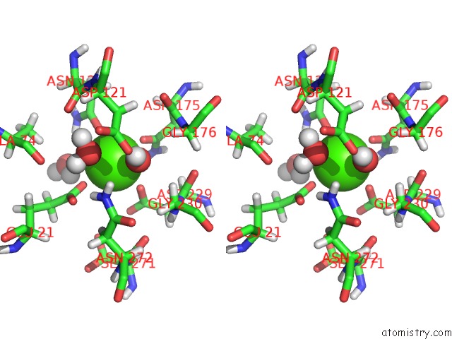
Stereo pair view
Mono view
Stereo pair view
A full contact list of Calcium with other atoms in the Ca binding
site number 1 of Dfpase at 0.85 Angstrom Resolution (H Atoms Included) within 5.0Å range:
|
Calcium binding site 2 out of 2 in 3o4p
Go back to
Calcium binding site 2 out
of 2 in the Dfpase at 0.85 Angstrom Resolution (H Atoms Included)
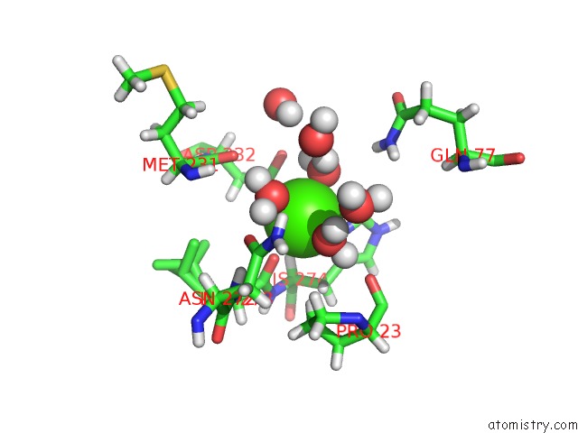
Mono view
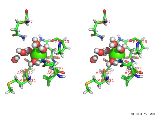
Stereo pair view
Mono view
Stereo pair view
A full contact list of Calcium with other atoms in the Ca binding
site number 2 of Dfpase at 0.85 Angstrom Resolution (H Atoms Included) within 5.0Å range:
|
Reference:
M.Elias,
D.Liebschner,
J.Koepke,
C.Lecomte,
B.Guillot,
C.Jelsch,
E.Chabriere.
Hydrogen Atoms in Protein Structures: High-Resolution X-Ray Diffraction Structure of the Dfpase. Bmc Res Notes V. 6 308 2013.
ISSN: ESSN 1756-0500
PubMed: 23915572
DOI: 10.1186/1756-0500-6-308
Page generated: Tue Jul 8 15:01:33 2025
ISSN: ESSN 1756-0500
PubMed: 23915572
DOI: 10.1186/1756-0500-6-308
Last articles
Cl in 5HGKCl in 5HGI
Cl in 5HG1
Cl in 5HGJ
Cl in 5HDO
Cl in 5HEZ
Cl in 5HG7
Cl in 5HFO
Cl in 5HES
Cl in 5HCJ