Calcium »
PDB 3nur-3ojn »
3o82 »
Calcium in PDB 3o82: Structure of Base N-Terminal Domain From Acinetobacter Baumannii Bound to 5'-O-[N-(2,3-Dihydroxybenzoyl)Sulfamoyl] Adenosine
Protein crystallography data
The structure of Structure of Base N-Terminal Domain From Acinetobacter Baumannii Bound to 5'-O-[N-(2,3-Dihydroxybenzoyl)Sulfamoyl] Adenosine, PDB code: 3o82
was solved by
E.J.Drake,
B.P.Duckworth,
J.Neres,
C.C.Aldrich,
A.M.Gulick,
with X-Ray Crystallography technique. A brief refinement statistics is given in the table below:
| Resolution Low / High (Å) | 40.00 / 2.70 |
| Space group | P 21 21 21 |
| Cell size a, b, c (Å), α, β, γ (°) | 65.447, 145.259, 149.063, 90.00, 90.00, 90.00 |
| R / Rfree (%) | 20 / 25.1 |
Calcium Binding Sites:
The binding sites of Calcium atom in the Structure of Base N-Terminal Domain From Acinetobacter Baumannii Bound to 5'-O-[N-(2,3-Dihydroxybenzoyl)Sulfamoyl] Adenosine
(pdb code 3o82). This binding sites where shown within
5.0 Angstroms radius around Calcium atom.
In total 4 binding sites of Calcium where determined in the Structure of Base N-Terminal Domain From Acinetobacter Baumannii Bound to 5'-O-[N-(2,3-Dihydroxybenzoyl)Sulfamoyl] Adenosine, PDB code: 3o82:
Jump to Calcium binding site number: 1; 2; 3; 4;
In total 4 binding sites of Calcium where determined in the Structure of Base N-Terminal Domain From Acinetobacter Baumannii Bound to 5'-O-[N-(2,3-Dihydroxybenzoyl)Sulfamoyl] Adenosine, PDB code: 3o82:
Jump to Calcium binding site number: 1; 2; 3; 4;
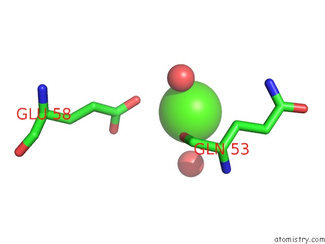
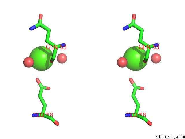
Calcium binding site 1 out of 4 in 3o82
Go back to
Calcium binding site 1 out
of 4 in the Structure of Base N-Terminal Domain From Acinetobacter Baumannii Bound to 5'-O-[N-(2,3-Dihydroxybenzoyl)Sulfamoyl] Adenosine
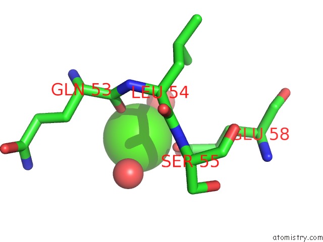
Mono view
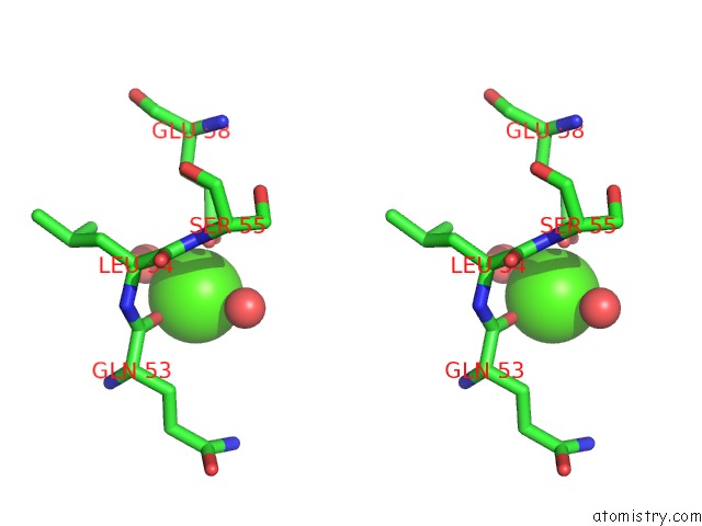
Stereo pair view
Mono view
Stereo pair view
A full contact list of Calcium with other atoms in the Ca binding
site number 1 of Structure of Base N-Terminal Domain From Acinetobacter Baumannii Bound to 5'-O-[N-(2,3-Dihydroxybenzoyl)Sulfamoyl] Adenosine within 5.0Å range:
|
Calcium binding site 2 out of 4 in 3o82
Go back to
Calcium binding site 2 out
of 4 in the Structure of Base N-Terminal Domain From Acinetobacter Baumannii Bound to 5'-O-[N-(2,3-Dihydroxybenzoyl)Sulfamoyl] Adenosine
Mono view
Stereo pair view
Mono view
Stereo pair view
A full contact list of Calcium with other atoms in the Ca binding
site number 2 of Structure of Base N-Terminal Domain From Acinetobacter Baumannii Bound to 5'-O-[N-(2,3-Dihydroxybenzoyl)Sulfamoyl] Adenosine within 5.0Å range:
|
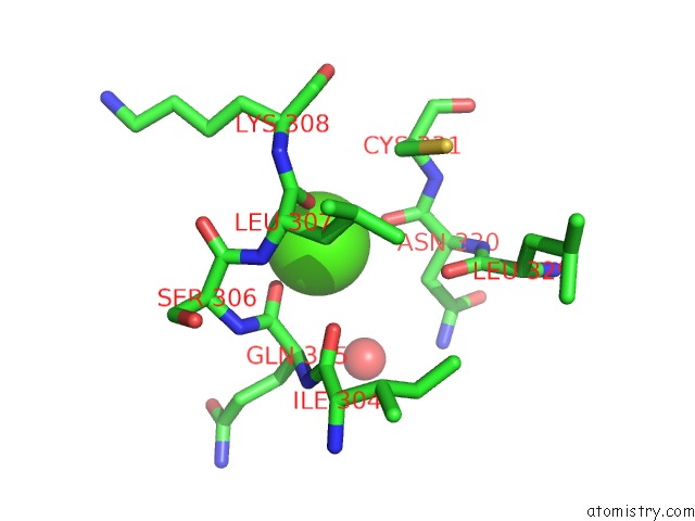
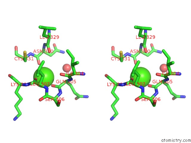
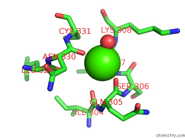
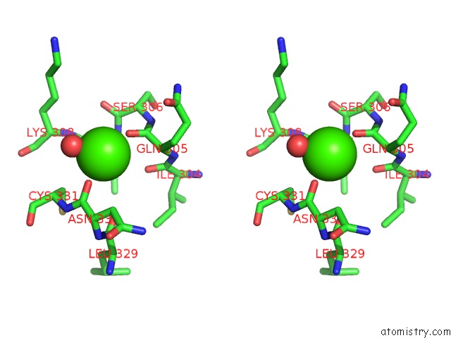
Calcium binding site 3 out of 4 in 3o82
Go back to
Calcium binding site 3 out
of 4 in the Structure of Base N-Terminal Domain From Acinetobacter Baumannii Bound to 5'-O-[N-(2,3-Dihydroxybenzoyl)Sulfamoyl] Adenosine
Mono view
Stereo pair view
Mono view
Stereo pair view
A full contact list of Calcium with other atoms in the Ca binding
site number 3 of Structure of Base N-Terminal Domain From Acinetobacter Baumannii Bound to 5'-O-[N-(2,3-Dihydroxybenzoyl)Sulfamoyl] Adenosine within 5.0Å range:
|
Calcium binding site 4 out of 4 in 3o82
Go back to
Calcium binding site 4 out
of 4 in the Structure of Base N-Terminal Domain From Acinetobacter Baumannii Bound to 5'-O-[N-(2,3-Dihydroxybenzoyl)Sulfamoyl] Adenosine
Mono view
Stereo pair view
Mono view
Stereo pair view
A full contact list of Calcium with other atoms in the Ca binding
site number 4 of Structure of Base N-Terminal Domain From Acinetobacter Baumannii Bound to 5'-O-[N-(2,3-Dihydroxybenzoyl)Sulfamoyl] Adenosine within 5.0Å range:
|
Reference:
E.J.Drake,
B.P.Duckworth,
J.Neres,
C.C.Aldrich,
A.M.Gulick.
Biochemical and Structural Characterization of Bisubstrate Inhibitors of Base, the Self-Standing Nonribosomal Peptide Synthetase Adenylate-Forming Enzyme of Acinetobactin Synthesis. Biochemistry V. 49 9292 2010.
ISSN: ISSN 0006-2960
PubMed: 20853905
DOI: 10.1021/BI101226N
Page generated: Tue Jul 8 15:03:11 2025
ISSN: ISSN 0006-2960
PubMed: 20853905
DOI: 10.1021/BI101226N
Last articles
Cl in 5JY1Cl in 5JYL
Cl in 5JXK
Cl in 5JXR
Cl in 5JXP
Cl in 5JXQ
Cl in 5JXJ
Cl in 5JX0
Cl in 5JXF
Cl in 5JX3