Calcium »
PDB 3nur-3ojn »
3ojk »
Calcium in PDB 3ojk: Structure of Co-Substituted Homoprotocatechuate 2,3-Dioxygenase in Complex with 4-Nitrocatechol at 1.68 Ang Resolution
Enzymatic activity of Structure of Co-Substituted Homoprotocatechuate 2,3-Dioxygenase in Complex with 4-Nitrocatechol at 1.68 Ang Resolution
All present enzymatic activity of Structure of Co-Substituted Homoprotocatechuate 2,3-Dioxygenase in Complex with 4-Nitrocatechol at 1.68 Ang Resolution:
1.13.11.15;
1.13.11.15;
Protein crystallography data
The structure of Structure of Co-Substituted Homoprotocatechuate 2,3-Dioxygenase in Complex with 4-Nitrocatechol at 1.68 Ang Resolution, PDB code: 3ojk
was solved by
A.J.Fielding,
E.G.Kovaleva,
E.R.Farquhar,
J.D.Lipscomb,
L.Que Jr.,
with X-Ray Crystallography technique. A brief refinement statistics is given in the table below:
| Resolution Low / High (Å) | 37.96 / 1.68 |
| Space group | P 21 21 2 |
| Cell size a, b, c (Å), α, β, γ (°) | 110.102, 150.395, 95.941, 90.00, 90.00, 90.00 |
| R / Rfree (%) | 15.3 / 18.2 |
Other elements in 3ojk:
The structure of Structure of Co-Substituted Homoprotocatechuate 2,3-Dioxygenase in Complex with 4-Nitrocatechol at 1.68 Ang Resolution also contains other interesting chemical elements:
| Cobalt | (Co) | 4 atoms |
| Chlorine | (Cl) | 4 atoms |
Calcium Binding Sites:
The binding sites of Calcium atom in the Structure of Co-Substituted Homoprotocatechuate 2,3-Dioxygenase in Complex with 4-Nitrocatechol at 1.68 Ang Resolution
(pdb code 3ojk). This binding sites where shown within
5.0 Angstroms radius around Calcium atom.
In total only one binding site of Calcium was determined in the Structure of Co-Substituted Homoprotocatechuate 2,3-Dioxygenase in Complex with 4-Nitrocatechol at 1.68 Ang Resolution, PDB code: 3ojk:
In total only one binding site of Calcium was determined in the Structure of Co-Substituted Homoprotocatechuate 2,3-Dioxygenase in Complex with 4-Nitrocatechol at 1.68 Ang Resolution, PDB code: 3ojk:
Calcium binding site 1 out of 1 in 3ojk
Go back to
Calcium binding site 1 out
of 1 in the Structure of Co-Substituted Homoprotocatechuate 2,3-Dioxygenase in Complex with 4-Nitrocatechol at 1.68 Ang Resolution
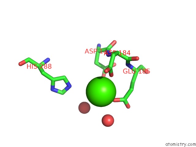
Mono view
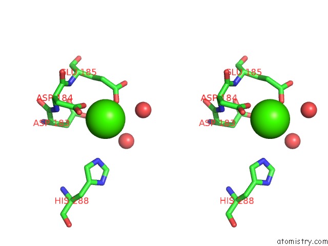
Stereo pair view
Mono view
Stereo pair view
A full contact list of Calcium with other atoms in the Ca binding
site number 1 of Structure of Co-Substituted Homoprotocatechuate 2,3-Dioxygenase in Complex with 4-Nitrocatechol at 1.68 Ang Resolution within 5.0Å range:
|
Reference:
A.J.Fielding,
E.G.Kovaleva,
E.R.Farquhar,
J.D.Lipscomb,
L.Que.
A Hyperactive Cobalt-Substituted Extradiol-Cleaving Catechol Dioxygenase. J.Biol.Inorg.Chem. V. 16 341 2011.
ISSN: ISSN 0949-8257
PubMed: 21153851
DOI: 10.1007/S00775-010-0732-0
Page generated: Tue Jul 8 15:06:30 2025
ISSN: ISSN 0949-8257
PubMed: 21153851
DOI: 10.1007/S00775-010-0732-0
Last articles
F in 4HXNF in 4HT2
F in 4HU1
F in 4HVS
F in 4HW7
F in 4HUA
F in 4HU9
F in 4HQJ
F in 4HT3
F in 4HLQ