Calcium »
PDB 3ojt-3p1h »
3ool »
Calcium in PDB 3ool: I-Scei Complexed with C/G+4 Dna Substrate
Protein crystallography data
The structure of I-Scei Complexed with C/G+4 Dna Substrate, PDB code: 3ool
was solved by
R.Joshi,
J.-H.Chen,
B.L.Golden,
F.S.Gimble,
with X-Ray Crystallography technique. A brief refinement statistics is given in the table below:
| Resolution Low / High (Å) | 30.79 / 2.30 |
| Space group | C 1 2 1 |
| Cell size a, b, c (Å), α, β, γ (°) | 129.184, 49.938, 63.360, 90.00, 107.54, 90.00 |
| R / Rfree (%) | 24.6 / 29.4 |
Calcium Binding Sites:
The binding sites of Calcium atom in the I-Scei Complexed with C/G+4 Dna Substrate
(pdb code 3ool). This binding sites where shown within
5.0 Angstroms radius around Calcium atom.
In total 3 binding sites of Calcium where determined in the I-Scei Complexed with C/G+4 Dna Substrate, PDB code: 3ool:
Jump to Calcium binding site number: 1; 2; 3;
In total 3 binding sites of Calcium where determined in the I-Scei Complexed with C/G+4 Dna Substrate, PDB code: 3ool:
Jump to Calcium binding site number: 1; 2; 3;
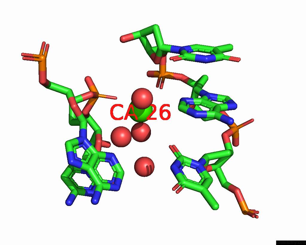
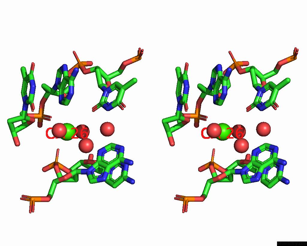
Calcium binding site 1 out of 3 in 3ool
Go back to
Calcium binding site 1 out
of 3 in the I-Scei Complexed with C/G+4 Dna Substrate
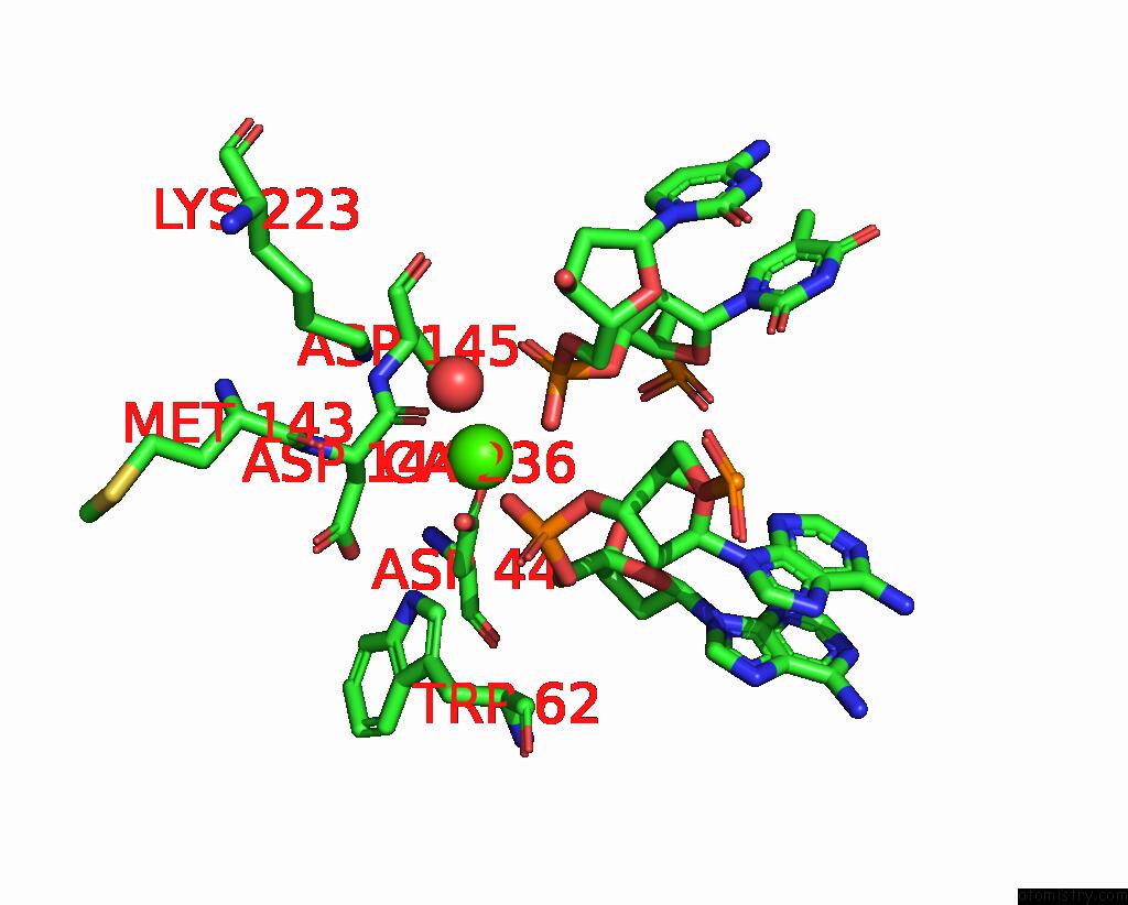
Mono view
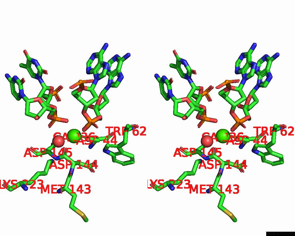
Stereo pair view
Mono view
Stereo pair view
A full contact list of Calcium with other atoms in the Ca binding
site number 1 of I-Scei Complexed with C/G+4 Dna Substrate within 5.0Å range:
|
Calcium binding site 2 out of 3 in 3ool
Go back to
Calcium binding site 2 out
of 3 in the I-Scei Complexed with C/G+4 Dna Substrate
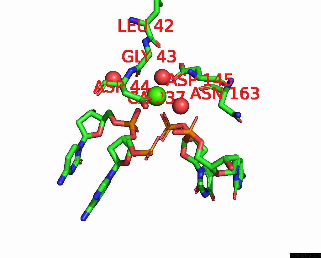
Mono view
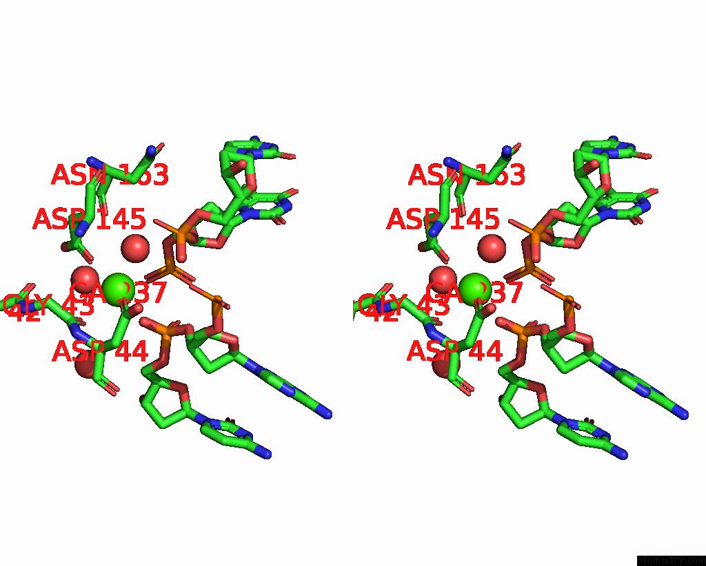
Stereo pair view
Mono view
Stereo pair view
A full contact list of Calcium with other atoms in the Ca binding
site number 2 of I-Scei Complexed with C/G+4 Dna Substrate within 5.0Å range:
|
Calcium binding site 3 out of 3 in 3ool
Go back to
Calcium binding site 3 out
of 3 in the I-Scei Complexed with C/G+4 Dna Substrate
Mono view
Stereo pair view
Mono view
Stereo pair view
A full contact list of Calcium with other atoms in the Ca binding
site number 3 of I-Scei Complexed with C/G+4 Dna Substrate within 5.0Å range:
|
Reference:
R.Joshi,
K.K.Ho,
K.Tenney,
J.H.Chen,
B.L.Golden,
F.S.Gimble.
Evolution of I-Scei Homing Endonucleases with Increased Dna Recognition Site Specificity. J.Mol.Biol. V. 405 185 2011.
ISSN: ISSN 0022-2836
PubMed: 21029741
DOI: 10.1016/J.JMB.2010.10.029
Page generated: Tue Jul 8 15:09:07 2025
ISSN: ISSN 0022-2836
PubMed: 21029741
DOI: 10.1016/J.JMB.2010.10.029
Last articles
Cl in 5II3Cl in 5IFM
Cl in 5IJ6
Cl in 5IHG
Cl in 5IHR
Cl in 5IGN
Cl in 5IH5
Cl in 5IG6
Cl in 5IFU
Cl in 5IFT