Calcium »
PDB 3ojt-3p1h »
3orv »
Calcium in PDB 3orv: Crystal Structure of the Y294H-Maug/Pre-Methylamine Dehydrogenase Complex
Enzymatic activity of Crystal Structure of the Y294H-Maug/Pre-Methylamine Dehydrogenase Complex
All present enzymatic activity of Crystal Structure of the Y294H-Maug/Pre-Methylamine Dehydrogenase Complex:
1.4.99.3;
1.4.99.3;
Protein crystallography data
The structure of Crystal Structure of the Y294H-Maug/Pre-Methylamine Dehydrogenase Complex, PDB code: 3orv
was solved by
L.M.R.Jensen,
C.M.Wilmot,
with X-Ray Crystallography technique. A brief refinement statistics is given in the table below:
| Resolution Low / High (Å) | 44.49 / 1.91 |
| Space group | P 1 |
| Cell size a, b, c (Å), α, β, γ (°) | 55.527, 83.524, 107.782, 109.94, 91.54, 105.78 |
| R / Rfree (%) | 13.9 / 18.7 |
Other elements in 3orv:
The structure of Crystal Structure of the Y294H-Maug/Pre-Methylamine Dehydrogenase Complex also contains other interesting chemical elements:
| Iron | (Fe) | 4 atoms |
Calcium Binding Sites:
The binding sites of Calcium atom in the Crystal Structure of the Y294H-Maug/Pre-Methylamine Dehydrogenase Complex
(pdb code 3orv). This binding sites where shown within
5.0 Angstroms radius around Calcium atom.
In total 2 binding sites of Calcium where determined in the Crystal Structure of the Y294H-Maug/Pre-Methylamine Dehydrogenase Complex, PDB code: 3orv:
Jump to Calcium binding site number: 1; 2;
In total 2 binding sites of Calcium where determined in the Crystal Structure of the Y294H-Maug/Pre-Methylamine Dehydrogenase Complex, PDB code: 3orv:
Jump to Calcium binding site number: 1; 2;
Calcium binding site 1 out of 2 in 3orv
Go back to
Calcium binding site 1 out
of 2 in the Crystal Structure of the Y294H-Maug/Pre-Methylamine Dehydrogenase Complex
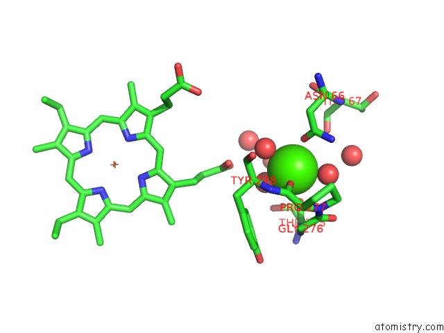
Mono view
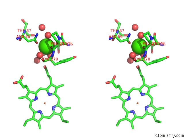
Stereo pair view
Mono view
Stereo pair view
A full contact list of Calcium with other atoms in the Ca binding
site number 1 of Crystal Structure of the Y294H-Maug/Pre-Methylamine Dehydrogenase Complex within 5.0Å range:
|
Calcium binding site 2 out of 2 in 3orv
Go back to
Calcium binding site 2 out
of 2 in the Crystal Structure of the Y294H-Maug/Pre-Methylamine Dehydrogenase Complex
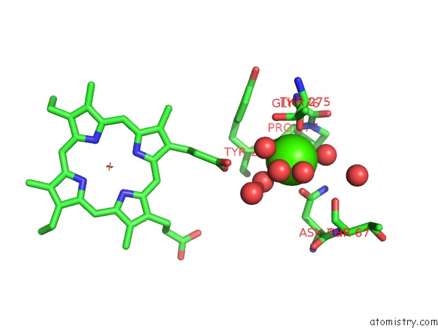
Mono view
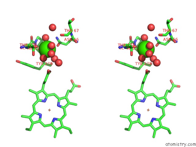
Stereo pair view
Mono view
Stereo pair view
A full contact list of Calcium with other atoms in the Ca binding
site number 2 of Crystal Structure of the Y294H-Maug/Pre-Methylamine Dehydrogenase Complex within 5.0Å range:
|
Reference:
N.Abu Tarboush,
L.M.Jensen,
M.Feng,
H.Tachikawa,
C.M.Wilmot,
V.L.Davidson.
Functional Importance of Tyrosine 294 and the Catalytic Selectivity For the Bis-Fe(IV) State of Maug Revealed By Replacement of This Axial Heme Ligand with Histidine . Biochemistry V. 49 9783 2010.
ISSN: ISSN 0006-2960
PubMed: 20929212
DOI: 10.1021/BI101254P
Page generated: Sat Jul 13 16:21:00 2024
ISSN: ISSN 0006-2960
PubMed: 20929212
DOI: 10.1021/BI101254P
Last articles
Zn in 9J0NZn in 9J0O
Zn in 9J0P
Zn in 9FJX
Zn in 9EKB
Zn in 9C0F
Zn in 9CAH
Zn in 9CH0
Zn in 9CH3
Zn in 9CH1