Calcium »
PDB 3ojt-3p1h »
3oss »
Calcium in PDB 3oss: The Crystal Structure of Enterotoxigenic Escherichia Coli Gspc-Gspd Complex From the Type II Secretion System
Protein crystallography data
The structure of The Crystal Structure of Enterotoxigenic Escherichia Coli Gspc-Gspd Complex From the Type II Secretion System, PDB code: 3oss
was solved by
K.V.Korotkov,
J.Pruneda,
W.G.J.Hol,
with X-Ray Crystallography technique. A brief refinement statistics is given in the table below:
| Resolution Low / High (Å) | 42.88 / 2.63 |
| Space group | P 21 21 21 |
| Cell size a, b, c (Å), α, β, γ (°) | 45.500, 76.810, 85.770, 90.00, 90.00, 90.00 |
| R / Rfree (%) | 21.3 / 26.5 |
Other elements in 3oss:
The structure of The Crystal Structure of Enterotoxigenic Escherichia Coli Gspc-Gspd Complex From the Type II Secretion System also contains other interesting chemical elements:
| Chlorine | (Cl) | 1 atom |
Calcium Binding Sites:
The binding sites of Calcium atom in the The Crystal Structure of Enterotoxigenic Escherichia Coli Gspc-Gspd Complex From the Type II Secretion System
(pdb code 3oss). This binding sites where shown within
5.0 Angstroms radius around Calcium atom.
In total only one binding site of Calcium was determined in the The Crystal Structure of Enterotoxigenic Escherichia Coli Gspc-Gspd Complex From the Type II Secretion System, PDB code: 3oss:
In total only one binding site of Calcium was determined in the The Crystal Structure of Enterotoxigenic Escherichia Coli Gspc-Gspd Complex From the Type II Secretion System, PDB code: 3oss:
Calcium binding site 1 out of 1 in 3oss
Go back to
Calcium binding site 1 out
of 1 in the The Crystal Structure of Enterotoxigenic Escherichia Coli Gspc-Gspd Complex From the Type II Secretion System
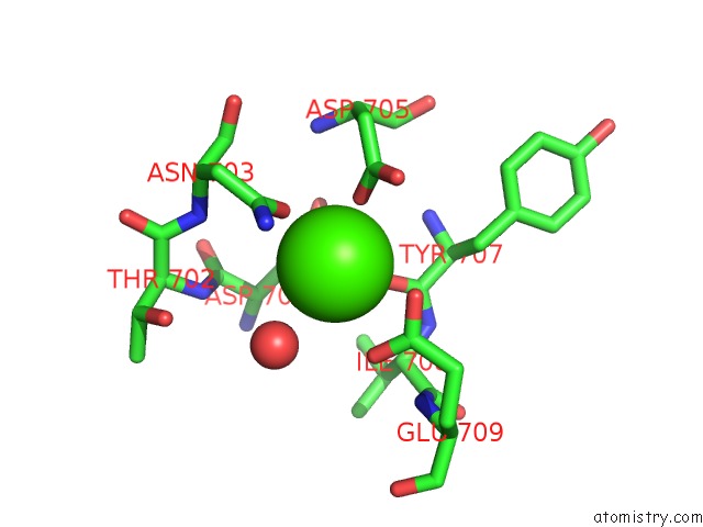
Mono view
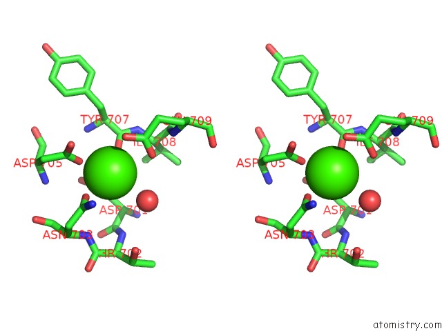
Stereo pair view
Mono view
Stereo pair view
A full contact list of Calcium with other atoms in the Ca binding
site number 1 of The Crystal Structure of Enterotoxigenic Escherichia Coli Gspc-Gspd Complex From the Type II Secretion System within 5.0Å range:
|
Reference:
K.V.Korotkov,
T.L.Johnson,
M.G.Jobling,
J.Pruneda,
E.Pardon,
A.Heroux,
S.Turley,
J.Steyaert,
R.K.Holmes,
M.Sandkvist,
W.G.Hol.
Structural and Functional Studies on the Interaction of Gspc and Gspd in the Type II Secretion System. Plos Pathog. V. 7 02228 2011.
ISSN: ISSN 1553-7366
PubMed: 21931548
DOI: 10.1371/JOURNAL.PPAT.1002228
Page generated: Sat Jul 13 16:21:55 2024
ISSN: ISSN 1553-7366
PubMed: 21931548
DOI: 10.1371/JOURNAL.PPAT.1002228
Last articles
Zn in 9J0NZn in 9J0O
Zn in 9J0P
Zn in 9FJX
Zn in 9EKB
Zn in 9C0F
Zn in 9CAH
Zn in 9CH0
Zn in 9CH3
Zn in 9CH1