Calcium »
PDB 3ojt-3p1h »
3otj »
Calcium in PDB 3otj: A Crystal Structure of Trypsin Complexed with Bpti (Bovine Pancreatic Trypsin Inhibitor) By X-Ray/Neutron Joint Refinement
Enzymatic activity of A Crystal Structure of Trypsin Complexed with Bpti (Bovine Pancreatic Trypsin Inhibitor) By X-Ray/Neutron Joint Refinement
All present enzymatic activity of A Crystal Structure of Trypsin Complexed with Bpti (Bovine Pancreatic Trypsin Inhibitor) By X-Ray/Neutron Joint Refinement:
3.4.21.4;
3.4.21.4;
Protein crystallography data
The structure of A Crystal Structure of Trypsin Complexed with Bpti (Bovine Pancreatic Trypsin Inhibitor) By X-Ray/Neutron Joint Refinement, PDB code: 3otj
was solved by
K.Kawamura,
T.Yamada,
K.Kurihara,
T.Tamada,
R.Kuroki,
I.Tanaka,
H.Takahashi,
N.Niimura,
with X-Ray Crystallography technique. A brief refinement statistics is given in the table below:
| Resolution Low / High (Å) | N/A / 2.15 |
| Space group | I 2 2 2 |
| Cell size a, b, c (Å), α, β, γ (°) | 75.604, 85.361, 122.551, 90.00, 90.00, 90.00 |
| R / Rfree (%) | 19.8 / 20.9 |
Calcium Binding Sites:
The binding sites of Calcium atom in the A Crystal Structure of Trypsin Complexed with Bpti (Bovine Pancreatic Trypsin Inhibitor) By X-Ray/Neutron Joint Refinement
(pdb code 3otj). This binding sites where shown within
5.0 Angstroms radius around Calcium atom.
In total only one binding site of Calcium was determined in the A Crystal Structure of Trypsin Complexed with Bpti (Bovine Pancreatic Trypsin Inhibitor) By X-Ray/Neutron Joint Refinement, PDB code: 3otj:
In total only one binding site of Calcium was determined in the A Crystal Structure of Trypsin Complexed with Bpti (Bovine Pancreatic Trypsin Inhibitor) By X-Ray/Neutron Joint Refinement, PDB code: 3otj:
Calcium binding site 1 out of 1 in 3otj
Go back to
Calcium binding site 1 out
of 1 in the A Crystal Structure of Trypsin Complexed with Bpti (Bovine Pancreatic Trypsin Inhibitor) By X-Ray/Neutron Joint Refinement
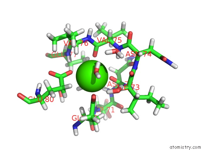
Mono view
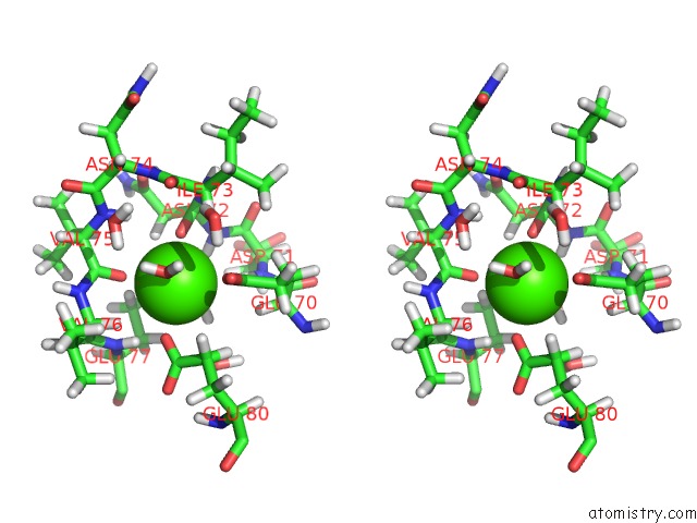
Stereo pair view
Mono view
Stereo pair view
A full contact list of Calcium with other atoms in the Ca binding
site number 1 of A Crystal Structure of Trypsin Complexed with Bpti (Bovine Pancreatic Trypsin Inhibitor) By X-Ray/Neutron Joint Refinement within 5.0Å range:
|
Reference:
K.Kawamura,
T.Yamada,
K.Kurihara,
T.Tamada,
R.Kuroki,
I.Tanaka,
H.Takahashi,
N.Niimura.
X-Ray and Neutron Protein Crystallographic Analysis of the Trypsin-Bpti Complex. Acta Crystallogr.,Sect.D V. 67 140 2011.
ISSN: ISSN 0907-4449
PubMed: 21245536
DOI: 10.1107/S0907444910053382
Page generated: Sat Jul 13 16:22:11 2024
ISSN: ISSN 0907-4449
PubMed: 21245536
DOI: 10.1107/S0907444910053382
Last articles
Zn in 9J0NZn in 9J0O
Zn in 9J0P
Zn in 9FJX
Zn in 9EKB
Zn in 9C0F
Zn in 9CAH
Zn in 9CH0
Zn in 9CH3
Zn in 9CH1