Calcium »
PDB 3p1o-3pdd »
3p5g »
Calcium in PDB 3p5g: Structure of the Carbohydrate-Recognition Domain of Human Langerin with Blood Group B Trisaccharide (Gal ALPHA1-3(Fuc ALPHA1-2)Gal)
Protein crystallography data
The structure of Structure of the Carbohydrate-Recognition Domain of Human Langerin with Blood Group B Trisaccharide (Gal ALPHA1-3(Fuc ALPHA1-2)Gal), PDB code: 3p5g
was solved by
H.Feinberg,
M.E.Taylor,
N.Razi,
R.Mcbride,
Y.A.Knirel,
S.A.Graham,
K.Drickamer,
W.I.Weis,
with X-Ray Crystallography technique. A brief refinement statistics is given in the table below:
| Resolution Low / High (Å) | 39.26 / 1.60 |
| Space group | P 42 |
| Cell size a, b, c (Å), α, β, γ (°) | 79.860, 79.860, 90.170, 90.00, 90.00, 90.00 |
| R / Rfree (%) | 17.9 / 21.9 |
Calcium Binding Sites:
The binding sites of Calcium atom in the Structure of the Carbohydrate-Recognition Domain of Human Langerin with Blood Group B Trisaccharide (Gal ALPHA1-3(Fuc ALPHA1-2)Gal)
(pdb code 3p5g). This binding sites where shown within
5.0 Angstroms radius around Calcium atom.
In total 4 binding sites of Calcium where determined in the Structure of the Carbohydrate-Recognition Domain of Human Langerin with Blood Group B Trisaccharide (Gal ALPHA1-3(Fuc ALPHA1-2)Gal), PDB code: 3p5g:
Jump to Calcium binding site number: 1; 2; 3; 4;
In total 4 binding sites of Calcium where determined in the Structure of the Carbohydrate-Recognition Domain of Human Langerin with Blood Group B Trisaccharide (Gal ALPHA1-3(Fuc ALPHA1-2)Gal), PDB code: 3p5g:
Jump to Calcium binding site number: 1; 2; 3; 4;
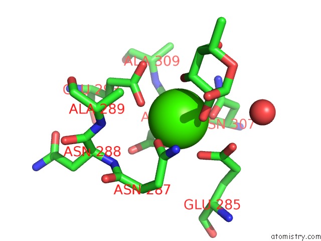
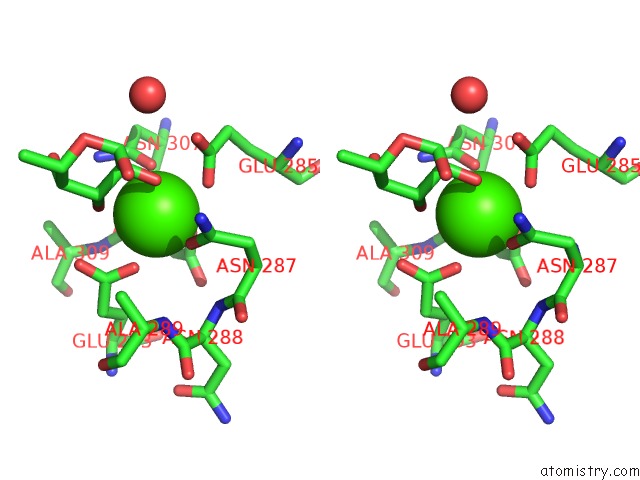
Calcium binding site 1 out of 4 in 3p5g
Go back to
Calcium binding site 1 out
of 4 in the Structure of the Carbohydrate-Recognition Domain of Human Langerin with Blood Group B Trisaccharide (Gal ALPHA1-3(Fuc ALPHA1-2)Gal)
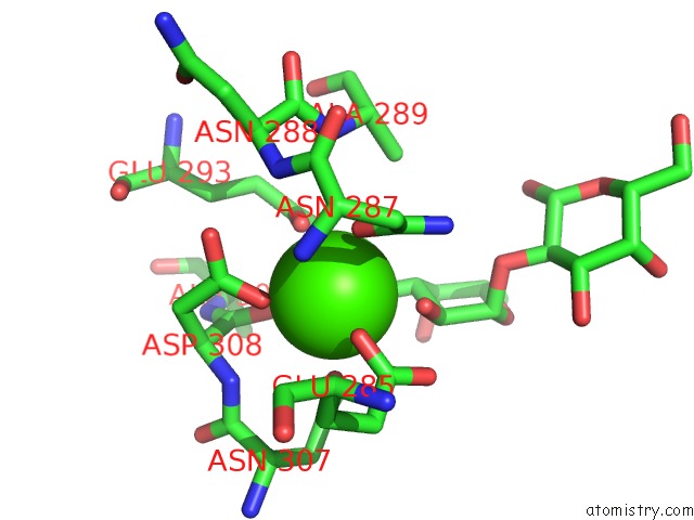
Mono view
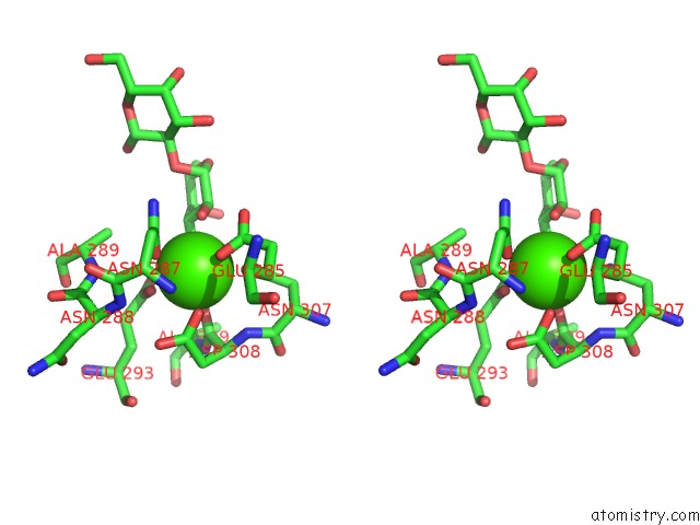
Stereo pair view
Mono view
Stereo pair view
A full contact list of Calcium with other atoms in the Ca binding
site number 1 of Structure of the Carbohydrate-Recognition Domain of Human Langerin with Blood Group B Trisaccharide (Gal ALPHA1-3(Fuc ALPHA1-2)Gal) within 5.0Å range:
|
Calcium binding site 2 out of 4 in 3p5g
Go back to
Calcium binding site 2 out
of 4 in the Structure of the Carbohydrate-Recognition Domain of Human Langerin with Blood Group B Trisaccharide (Gal ALPHA1-3(Fuc ALPHA1-2)Gal)
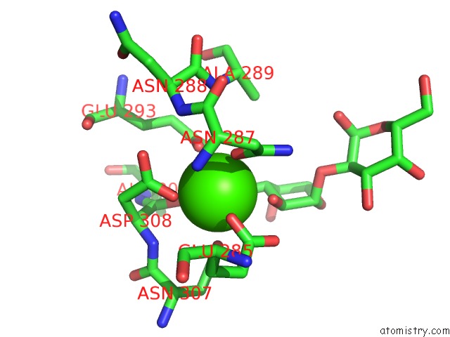
Mono view
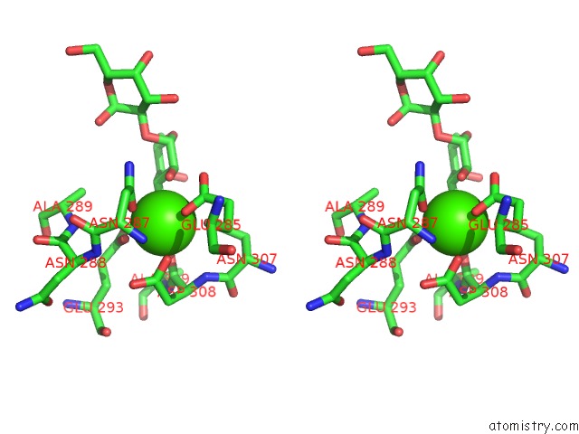
Stereo pair view
Mono view
Stereo pair view
A full contact list of Calcium with other atoms in the Ca binding
site number 2 of Structure of the Carbohydrate-Recognition Domain of Human Langerin with Blood Group B Trisaccharide (Gal ALPHA1-3(Fuc ALPHA1-2)Gal) within 5.0Å range:
|
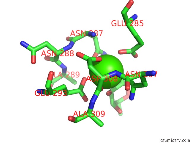
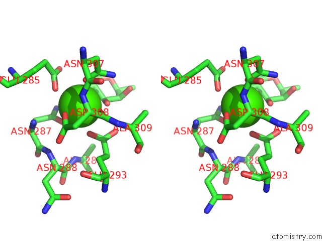
Calcium binding site 3 out of 4 in 3p5g
Go back to
Calcium binding site 3 out
of 4 in the Structure of the Carbohydrate-Recognition Domain of Human Langerin with Blood Group B Trisaccharide (Gal ALPHA1-3(Fuc ALPHA1-2)Gal)
Mono view
Stereo pair view
Mono view
Stereo pair view
A full contact list of Calcium with other atoms in the Ca binding
site number 3 of Structure of the Carbohydrate-Recognition Domain of Human Langerin with Blood Group B Trisaccharide (Gal ALPHA1-3(Fuc ALPHA1-2)Gal) within 5.0Å range:
|
Calcium binding site 4 out of 4 in 3p5g
Go back to
Calcium binding site 4 out
of 4 in the Structure of the Carbohydrate-Recognition Domain of Human Langerin with Blood Group B Trisaccharide (Gal ALPHA1-3(Fuc ALPHA1-2)Gal)
Mono view
Stereo pair view
Mono view
Stereo pair view
A full contact list of Calcium with other atoms in the Ca binding
site number 4 of Structure of the Carbohydrate-Recognition Domain of Human Langerin with Blood Group B Trisaccharide (Gal ALPHA1-3(Fuc ALPHA1-2)Gal) within 5.0Å range:
|
Reference:
H.Feinberg,
M.E.Taylor,
N.Razi,
R.Mcbride,
Y.A.Knirel,
S.A.Graham,
K.Drickamer,
W.I.Weis.
Structural Basis For Langerin Recognition of Diverse Pathogen and Mammalian Glycans Through A Single Binding Site. J.Mol.Biol. V. 405 1027 2011.
ISSN: ISSN 0022-2836
PubMed: 21112338
DOI: 10.1016/J.JMB.2010.11.039
Page generated: Sat Jul 13 16:31:34 2024
ISSN: ISSN 0022-2836
PubMed: 21112338
DOI: 10.1016/J.JMB.2010.11.039
Last articles
Zn in 9J0NZn in 9J0O
Zn in 9J0P
Zn in 9FJX
Zn in 9EKB
Zn in 9C0F
Zn in 9CAH
Zn in 9CH0
Zn in 9CH3
Zn in 9CH1