Calcium »
PDB 3p1o-3pdd »
3pak »
Calcium in PDB 3pak: Crystal Structure of Rat Surfactant Protein A Neck and Carbohydrate Recognition Domain (Ncrd) Complexed with Mannose
Protein crystallography data
The structure of Crystal Structure of Rat Surfactant Protein A Neck and Carbohydrate Recognition Domain (Ncrd) Complexed with Mannose, PDB code: 3pak
was solved by
F.Shang,
M.J.Rynkiewicz,
F.X.Mccormack,
H.Wu,
T.M.Cafarella,
J.Head,
B.A.Seaton,
with X-Ray Crystallography technique. A brief refinement statistics is given in the table below:
| Resolution Low / High (Å) | 26.10 / 1.90 |
| Space group | P 63 |
| Cell size a, b, c (Å), α, β, γ (°) | 97.796, 97.796, 45.088, 90.00, 90.00, 120.00 |
| R / Rfree (%) | 21.8 / 24.3 |
Other elements in 3pak:
The structure of Crystal Structure of Rat Surfactant Protein A Neck and Carbohydrate Recognition Domain (Ncrd) Complexed with Mannose also contains other interesting chemical elements:
| Sodium | (Na) | 1 atom |
Calcium Binding Sites:
The binding sites of Calcium atom in the Crystal Structure of Rat Surfactant Protein A Neck and Carbohydrate Recognition Domain (Ncrd) Complexed with Mannose
(pdb code 3pak). This binding sites where shown within
5.0 Angstroms radius around Calcium atom.
In total only one binding site of Calcium was determined in the Crystal Structure of Rat Surfactant Protein A Neck and Carbohydrate Recognition Domain (Ncrd) Complexed with Mannose, PDB code: 3pak:
In total only one binding site of Calcium was determined in the Crystal Structure of Rat Surfactant Protein A Neck and Carbohydrate Recognition Domain (Ncrd) Complexed with Mannose, PDB code: 3pak:
Calcium binding site 1 out of 1 in 3pak
Go back to
Calcium binding site 1 out
of 1 in the Crystal Structure of Rat Surfactant Protein A Neck and Carbohydrate Recognition Domain (Ncrd) Complexed with Mannose
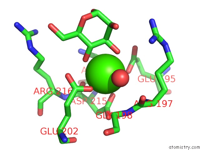
Mono view
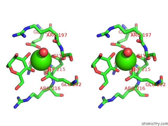
Stereo pair view
Mono view
Stereo pair view
A full contact list of Calcium with other atoms in the Ca binding
site number 1 of Crystal Structure of Rat Surfactant Protein A Neck and Carbohydrate Recognition Domain (Ncrd) Complexed with Mannose within 5.0Å range:
|
Reference:
F.Shang,
M.J.Rynkiewicz,
F.X.Mccormack,
H.Wu,
T.M.Cafarella,
J.F.Head,
B.A.Seaton.
Crystallographic Complexes of Surfactant Protein A and Carbohydrates Reveal Ligand-Induced Conformational Change. J.Biol.Chem. V. 286 757 2011.
ISSN: ISSN 0021-9258
PubMed: 21047777
DOI: 10.1074/JBC.M110.175265
Page generated: Tue Jul 8 15:25:07 2025
ISSN: ISSN 0021-9258
PubMed: 21047777
DOI: 10.1074/JBC.M110.175265
Last articles
Fe in 2YXOFe in 2YRS
Fe in 2YXC
Fe in 2YNM
Fe in 2YVJ
Fe in 2YP1
Fe in 2YU2
Fe in 2YU1
Fe in 2YQB
Fe in 2YOO