Calcium »
PDB 3p1o-3pdd »
3pat »
Calcium in PDB 3pat: Comparison Between the Crystal and the Solution Structures of the Ef Hand Parvalbumin
Calcium Binding Sites:
The binding sites of Calcium atom in the Comparison Between the Crystal and the Solution Structures of the Ef Hand Parvalbumin
(pdb code 3pat). This binding sites where shown within
5.0 Angstroms radius around Calcium atom.
In total 2 binding sites of Calcium where determined in the Comparison Between the Crystal and the Solution Structures of the Ef Hand Parvalbumin, PDB code: 3pat:
Jump to Calcium binding site number: 1; 2;
In total 2 binding sites of Calcium where determined in the Comparison Between the Crystal and the Solution Structures of the Ef Hand Parvalbumin, PDB code: 3pat:
Jump to Calcium binding site number: 1; 2;
Calcium binding site 1 out of 2 in 3pat
Go back to
Calcium binding site 1 out
of 2 in the Comparison Between the Crystal and the Solution Structures of the Ef Hand Parvalbumin
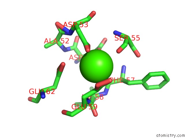
Mono view
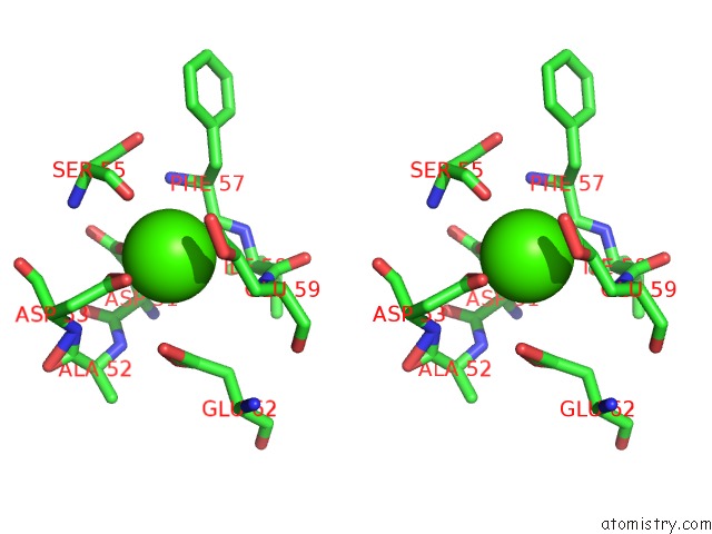
Stereo pair view
Mono view
Stereo pair view
A full contact list of Calcium with other atoms in the Ca binding
site number 1 of Comparison Between the Crystal and the Solution Structures of the Ef Hand Parvalbumin within 5.0Å range:
|
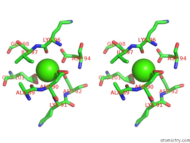
Calcium binding site 2 out of 2 in 3pat
Go back to
Calcium binding site 2 out
of 2 in the Comparison Between the Crystal and the Solution Structures of the Ef Hand Parvalbumin

Mono view
Stereo pair view
Mono view
Stereo pair view
A full contact list of Calcium with other atoms in the Ca binding
site number 2 of Comparison Between the Crystal and the Solution Structures of the Ef Hand Parvalbumin within 5.0Å range:
|
Reference:
A.Padilla,
A.Cave,
J.Parello,
G.Etienne,
C.Baldellon.
Comparison Between the Crystal and the Solution Structures of the Ef Hand Parvalbumin To Be Published.
Page generated: Sat Jul 13 16:40:51 2024
Last articles
Zn in 9J0NZn in 9J0O
Zn in 9J0P
Zn in 9FJX
Zn in 9EKB
Zn in 9C0F
Zn in 9CAH
Zn in 9CH0
Zn in 9CH3
Zn in 9CH1