Calcium »
PDB 3pe0-3pty »
3pe0 »
Calcium in PDB 3pe0: Structure of the Central Region of the Plakin Domain of Plectin
Protein crystallography data
The structure of Structure of the Central Region of the Plakin Domain of Plectin, PDB code: 3pe0
was solved by
E.Ortega,
J.M.De Pereda,
with X-Ray Crystallography technique. A brief refinement statistics is given in the table below:
| Resolution Low / High (Å) | 19.98 / 2.95 |
| Space group | P 21 21 21 |
| Cell size a, b, c (Å), α, β, γ (°) | 72.650, 108.512, 112.139, 90.00, 90.00, 90.00 |
| R / Rfree (%) | 24.1 / 26.5 |
Calcium Binding Sites:
The binding sites of Calcium atom in the Structure of the Central Region of the Plakin Domain of Plectin
(pdb code 3pe0). This binding sites where shown within
5.0 Angstroms radius around Calcium atom.
In total 2 binding sites of Calcium where determined in the Structure of the Central Region of the Plakin Domain of Plectin, PDB code: 3pe0:
Jump to Calcium binding site number: 1; 2;
In total 2 binding sites of Calcium where determined in the Structure of the Central Region of the Plakin Domain of Plectin, PDB code: 3pe0:
Jump to Calcium binding site number: 1; 2;
Calcium binding site 1 out of 2 in 3pe0
Go back to
Calcium binding site 1 out
of 2 in the Structure of the Central Region of the Plakin Domain of Plectin
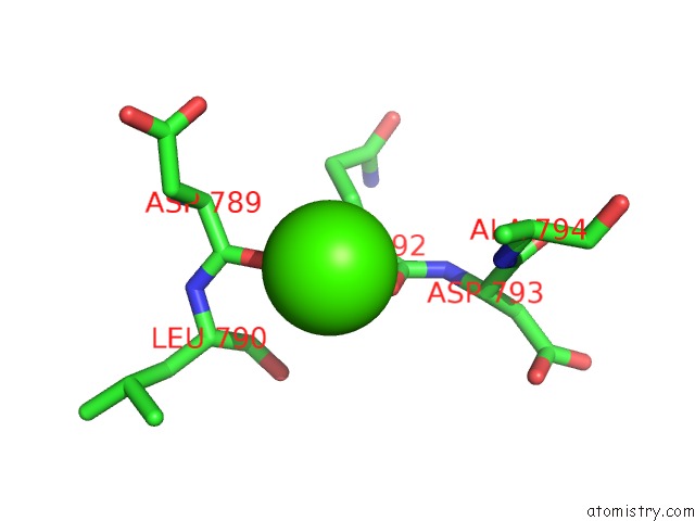
Mono view
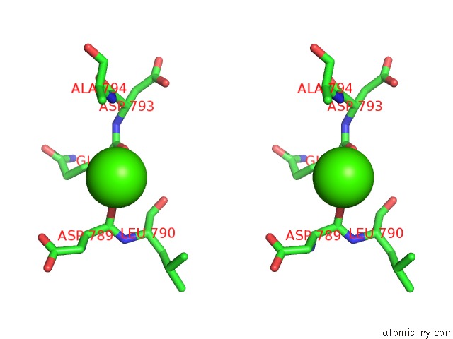
Stereo pair view
Mono view
Stereo pair view
A full contact list of Calcium with other atoms in the Ca binding
site number 1 of Structure of the Central Region of the Plakin Domain of Plectin within 5.0Å range:
|
Calcium binding site 2 out of 2 in 3pe0
Go back to
Calcium binding site 2 out
of 2 in the Structure of the Central Region of the Plakin Domain of Plectin

Mono view
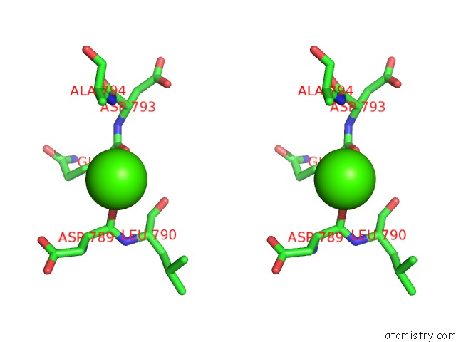
Stereo pair view
Mono view
Stereo pair view
A full contact list of Calcium with other atoms in the Ca binding
site number 2 of Structure of the Central Region of the Plakin Domain of Plectin within 5.0Å range:
|
Reference:
E.Ortega,
R.M.Buey,
A.Sonnenberg,
J.M.De Pereda.
The Structure of the Plakin Domain of Plectin Reveals A Non-Canonical SH3 Domain Interacting with Its Fourth Spectrin Repeat. J.Biol.Chem. V. 286 12429 2011.
ISSN: ISSN 0021-9258
PubMed: 21288893
DOI: 10.1074/JBC.M110.197467
Page generated: Sat Jul 13 16:49:22 2024
ISSN: ISSN 0021-9258
PubMed: 21288893
DOI: 10.1074/JBC.M110.197467
Last articles
Zn in 9J0NZn in 9J0O
Zn in 9J0P
Zn in 9FJX
Zn in 9EKB
Zn in 9C0F
Zn in 9CAH
Zn in 9CH0
Zn in 9CH3
Zn in 9CH1