Calcium »
PDB 3pe0-3pty »
3pog »
Calcium in PDB 3pog: Crystal Structure of the Masp-1 CUB2 Domain Bound to CA2+
Protein crystallography data
The structure of Crystal Structure of the Masp-1 CUB2 Domain Bound to CA2+, PDB code: 3pog
was solved by
A.R.Gingras,
P.C.E.Moody,
R.Wallis,
with X-Ray Crystallography technique. A brief refinement statistics is given in the table below:
| Resolution Low / High (Å) | 38.98 / 2.75 |
| Space group | C 1 2 1 |
| Cell size a, b, c (Å), α, β, γ (°) | 111.680, 64.690, 52.420, 90.00, 92.33, 90.00 |
| R / Rfree (%) | 23.3 / 28.6 |
Calcium Binding Sites:
The binding sites of Calcium atom in the Crystal Structure of the Masp-1 CUB2 Domain Bound to CA2+
(pdb code 3pog). This binding sites where shown within
5.0 Angstroms radius around Calcium atom.
In total 3 binding sites of Calcium where determined in the Crystal Structure of the Masp-1 CUB2 Domain Bound to CA2+, PDB code: 3pog:
Jump to Calcium binding site number: 1; 2; 3;
In total 3 binding sites of Calcium where determined in the Crystal Structure of the Masp-1 CUB2 Domain Bound to CA2+, PDB code: 3pog:
Jump to Calcium binding site number: 1; 2; 3;
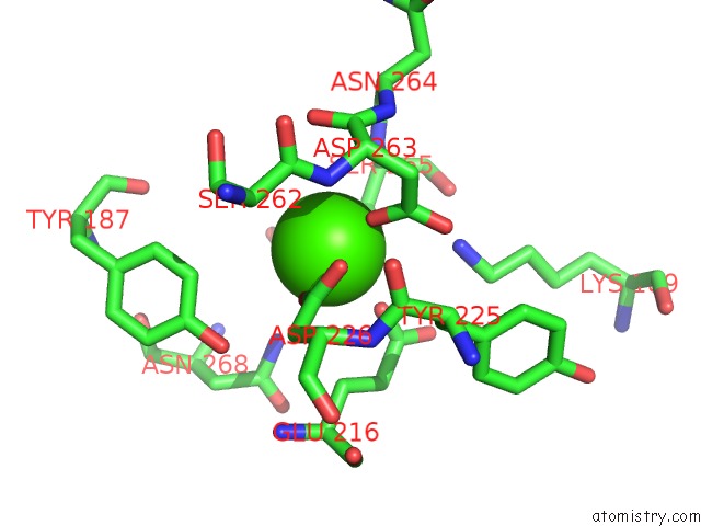
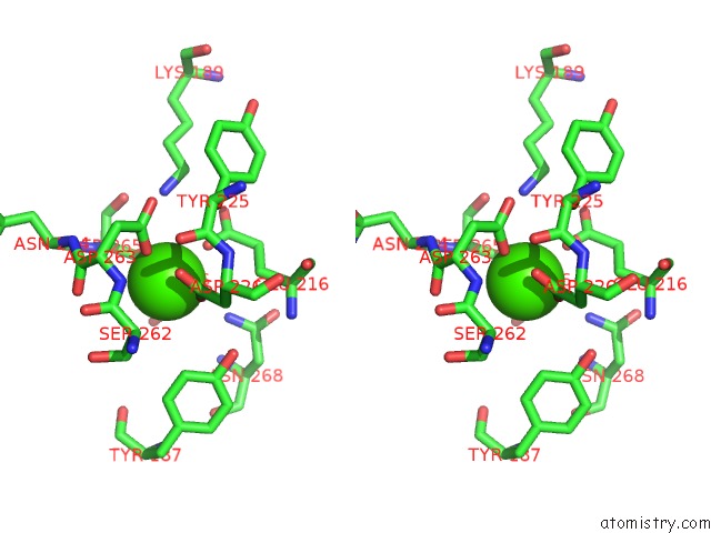
Calcium binding site 1 out of 3 in 3pog
Go back to
Calcium binding site 1 out
of 3 in the Crystal Structure of the Masp-1 CUB2 Domain Bound to CA2+
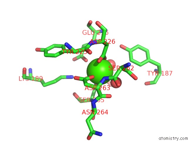
Mono view
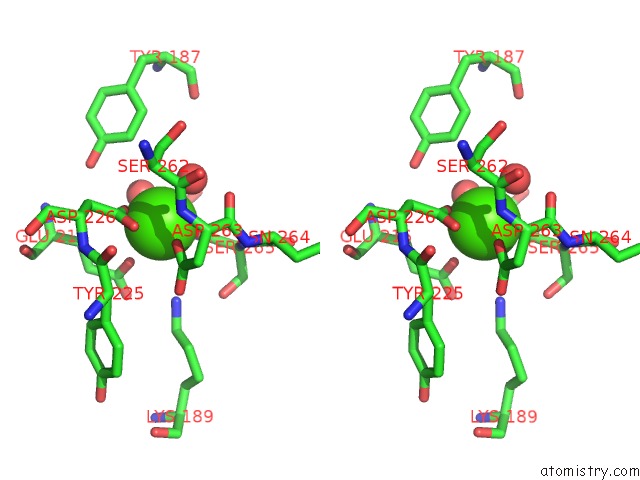
Stereo pair view
Mono view
Stereo pair view
A full contact list of Calcium with other atoms in the Ca binding
site number 1 of Crystal Structure of the Masp-1 CUB2 Domain Bound to CA2+ within 5.0Å range:
|
Calcium binding site 2 out of 3 in 3pog
Go back to
Calcium binding site 2 out
of 3 in the Crystal Structure of the Masp-1 CUB2 Domain Bound to CA2+
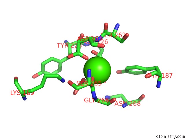
Mono view
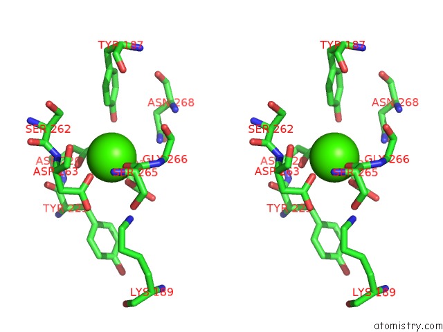
Stereo pair view
Mono view
Stereo pair view
A full contact list of Calcium with other atoms in the Ca binding
site number 2 of Crystal Structure of the Masp-1 CUB2 Domain Bound to CA2+ within 5.0Å range:
|
Calcium binding site 3 out of 3 in 3pog
Go back to
Calcium binding site 3 out
of 3 in the Crystal Structure of the Masp-1 CUB2 Domain Bound to CA2+
Mono view
Stereo pair view
Mono view
Stereo pair view
A full contact list of Calcium with other atoms in the Ca binding
site number 3 of Crystal Structure of the Masp-1 CUB2 Domain Bound to CA2+ within 5.0Å range:
|
Reference:
A.R.Gingras,
U.V.Girija,
A.H.Keeble,
R.Panchal,
D.A.Mitchell,
P.C.Moody,
R.Wallis.
Structural Basis of Mannan-Binding Lectin Recognition By Its Associated Serine Protease Masp-1: Implications For Complement Activation. Structure V. 19 1635 2011.
ISSN: ISSN 0969-2126
PubMed: 22078562
DOI: 10.1016/J.STR.2011.08.014
Page generated: Sat Jul 13 16:52:56 2024
ISSN: ISSN 0969-2126
PubMed: 22078562
DOI: 10.1016/J.STR.2011.08.014
Last articles
Zn in 9J0NZn in 9J0O
Zn in 9J0P
Zn in 9FJX
Zn in 9EKB
Zn in 9C0F
Zn in 9CAH
Zn in 9CH0
Zn in 9CH3
Zn in 9CH1